Wednesday, 16 February 2011 21:22

Tendons

Rate this item
(0 votes)

The deformation that occurs as force is applied and removed is called “elastic” deformation. The deformation that occurs after force application or removal is called “viscous” deformation. Because tissues of the body exhibit both elastic and viscous properties, they are called “viscoelastic”. If the recovery time between successive exertions is not long enough for a given force and duration, the recovery will not be complete and the tendon will be stretched further with each successive exertion. Goldstein et al. (1987) found that when finger flexor tendons were subjected to 8 seconds (s) physiological loads and 2 s rest, the accumulated viscous strain after 500 cycles was equal to the elastic strain. When the tendons were subjected to 2 s work and 8 s rest, the accumulated viscous strain after 500 cycles was negligible. Critical recovery times for given work-rest profiles have not yet been determined.

Tendons can be characterized as composite structures with parallel bundles of collagen fibres arranged in a gelatinous matrix of mucopolysaccharide. Tensile forces on the ends of the tendon cause unfolding of corrugations and straightening of the collagen strands. Additional loads cause stretching of the straightened strands. Consequently, the tendon gets stiffer as it gets longer. Compressive forces perpendicular to the long axis of the tendon cause the collagen strands to be forced closer together, and result in a flattening of the tendon. Shear forces on the side of the tendon cause displacement of the collagen strands closest to the surface with respect to those farthest away, and gives the side view of the tendon a skewed look.

Tendons as Structures

Forces are transmitted through tendons to maintain static and dynamic balance for specified work requirements. Contracting muscles tend to rotate the joints in one direction while the weight of the body and of work objects tends to rotate them in the other. Exact determination of these tendon forces is not possible because there are multiple muscles and tendons acting about each joint structure; however, it can be shown that the muscle forces acting on the tendons are much greater than the weight or reaction forces of work objects.

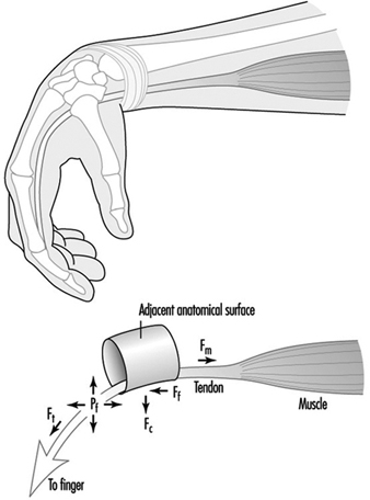
The forces exerted by contracting muscles are called tensile forces because they stretch the tendon. Tensile forces can be demonstrated by pulling on the ends of a rubber band. Tendons also are subjected to compressive and shear forces and to fluid pressures, which are illustrated in Figure 4 for the finger flexor tendons in the wrist.

Figure 1. Schematic diagram of tendon stretched around an anatomical surface or pulley and the corresponding tensile forces (Ft), compressive forces (Fc), friction forces (Ff) and hydrostatic or fluid pressure (Pf).

MUS040F1

Exertion of the fingers to grasp or manipulate work objects requires the contraction of muscles in the forearm and hand. As the muscles contract, they pull on the ends of their respective tendons, which pass through the centre and circumference of the wrist. If the wrist is not held in a position so that the tendons are perfectly straight, they will press against adjacent structures. The finger flexor tendons press against the bones and ligaments inside the carpal tunnel. These tendons can be seen to protrude under the skin toward the palm during forceful pinching with a flexed wrist. Similarly, the extensor and abductor tendons can be seen to protrude on the back and side of the wrist when it is extended with outstretched fingers.

Friction or shear forces are caused by dynamic exertions in which the tendons rub against adjacent anatomical surfaces. These forces act on and parallel to the surface of the tendon. Friction forces can be felt by simultaneously pressing and sliding the hand against a flat surface. The sliding of tendons over an adjacent anatomical surface is analogous to a belt sliding around a pulley.

Fluid pressure is caused by exertions or postures that displace fluid out of the spaces around the tendons. Studies of carpal canal pressure show that wrist contact with external surfaces                                                                                                                           and certain postures produce pressures high enough to impair                                                                                                                       circulation and threaten tissue viability (Lundborg 1988).

Contraction of a muscle produces an immediate stretching of its tendon. Tendons join muscles together. If the exertion is sustained, the tendon will continue to stretch. Relaxation of the muscle will result in a rapid recovery of the tendon followed by a slowed recovery. If the initial stretching was within certain limits, the tendon will recover to its initial unloaded length (Fung 1972).

Tendons as Living Tissues

The strength of tendons belies the delicacy of the underlying physiological mechanisms by which they are nourished and heal. Interspersed within the tendon matrix are living cells, nerve endings and blood vessels. Nerve endings provide information to the central nervous system for motor control and warning of acute overload. Blood vessels play an important role in the nourishment of some areas of the tendon. Some areas of tendons are avascular and rely on diffusion from fluid secreted by synovial linings of outer tendon sheaths (Gelberman et al. 1987). Synovial fluid also lubricates movements of the tendons. Synovial sheaths are found at locations where tendons come into contact with adjacent anatomical surfaces.

Excessive elastic or viscous deformation of the tendon can damage these tissues and impair their ability to heal. It is hypothesized that deformation may impede or arrest circulation and nourishment of tendons (Hagberg 1982; Viikari-Juntura 1984; Armstrong et al. 1993). Without adequate circulation, cell viability will be impaired and the tendon’s capacity to heal will be reduced. Tendon deformation can lead to small tears that further contribute to cell damage and inflammation. If circulation is restored and the tendon is given adequate recovery time, the damaged tissues will heal (Gelberman et al. 1987; Daniel and Breidenbach 1982; Leadbetter 1989).

Tendon Disorders

It has been shown that tendon disorders occur in predictable patterns (Armstrong et al. 1993). Their locations occur in those parts of the body associated with high stress concentrations (e.g., in the tendons of the supraspinatus, the biceps, the extrinsic finger flexor and extensor muscles). Also, there is an association between the intensity of work and the prevalence of tendon disorders. This pattern also has been shown for amateur and professional athletes (Leadbetter 1989). The common factors in both workers and athletes are repetitive exertions and overloading of the muscle-tendon units.

Within certain limits, the injuries produced by mechanical loading will heal. The healing process is divided into three stages: inflammatory, proliferatory and remodelling (Gelberman et al. 1987; Daniel and Breidenbach 1982). The inflammatory stage is characterized by the presence of polymorphonuclear cell infilt- ration, capillary budding and exudation, and lasts for several days. The proliferatory stage is characterized by the proliferation of fibroblasts and randomly oriented collagen fibres between areas of the wound and adjacent tissues, and lasts for several weeks. The remodelling phase is characterized by the alignment of the collagen fibres along the direction of loading, and lasts for several months. If the tissues are re-injured before healing is complete, recovery may be delayed and the condition may worsen (Leadbetter 1989). Normally healing leads to a strengthening or adaptation of the tissue to mechanical stress.

The effects of repetitive loading are apparent in the forearm finger flexor tendons where they contact the inside walls of the carpal tunnel (Louis 1992; Armstrong et al. 1984). It has been shown that there is progressive thickening of the synovial tissue between the edges of the carpal tunnel and the centre where the contact stresses on the tendons are the greatest. Thickening of the tendons is accompanied by synovial hyperplasia and proliferation of connective tissue. Thickening of the tendon sheaths is a widely cited factor in compression of the median nerve inside the carpal tunnel. It can be argued that thickening of the synovial tissues is an adaptation of the tendons to mechanical trauma. Were it not for the secondary effect on the median nerve compression resulting in carpal tunnel syndrome, it might be considered a desirable outcome.

Until optimal tendon loading regimes are determined, employers should monitor workers for signs or symptoms of tendon disorders so that they can intervene with work modifications to prevent further injuries. Jobs should be inspected for conspicuous risk factors any time an upper limb problem is identified or suspected. Jobs also should be inspected any time there is a change in the work standard, procedure or tooling, to insure that risk factors are minimized.

 

Back

Read 9176 times Last modified on Wednesday, 03 August 2011 21:11
More in this category: « Muscles Bones and Joints »

" DISCLAIMER: The ILO does not take responsibility for content presented on this web portal that is presented in any language other than English, which is the language used for the initial production and peer-review of original content. Certain statistics have not been updated since the production of the 4th edition of the Encyclopaedia (1998)."

Contents

Musculoskeletal System References

Agency for Health Care Policy and Research (AHCPR). 1994. Acute low-back problems in adults. Clinical Pratice Guidelines 14. Washington, DC: AHCPR.

Allander, E. 1974. Prevalence, incidence and remission rates of some common rheumatic diseases or syndromes. Scand J Rheumatol 3:145-153.

American Academy of Orthopaedic Surgeons. 1988. Joint Motion. New York: Churchill Livingstone.
Anderson, JAD. 1988. Arthrosis and its relation to work. Scand J Work Environ Health 10:429-433.

Anderson, JJ and DT Felson. 1988. Factors associated with osteoarthritis of the knee in the first National Health and Nutrition Survey (HANES 1): Evidence for an association with overweight, race and physical demands of work. Am J Epidemiol 128:179-189.

Angelides, AC. 1982. Ganglions of the hand and wrist. In Operative Hand Surgery, edited by DP Green. New York: Churchill Livingstone.

Armstrong, TJ, WA Castelli, G Evans, and R Diaz-Perez. 1984. Some histological changes in carpal tunnel contents and their biomechanical implications. J Occup Med 26(3):197-201.

Armstrong, TJ, P Buckle, L Fine, M Hagberg, B Jonsson, A Kilbom, I Kuorinka, B Silverstein, B Sjøgaard, and E Viikari-Juntura. 1993. A conceptual model for work-related neck and upper-limb musculoskeletal disorders. Scand J Work Environ Health 19:73-84.

Arnett, FC, SM Edworthy, DA Bloch, DJ McShane, JF Fries, NS Cooper, LA Healey, SR Kaplan, MH Liang, HS Luthra, TAJ Medsger, DM Mitchell, DH Neustadt, RS Pinals, JG Schaller, JT Sharp, RL Wilder, and GG Hunder. 1988. The American Rheumatism Association 1987 revised criteria for the classification of rheumatoid arthritis. Arthritis Rheum 31:315-324.

Aronsson, G, U Bergkvist, and S Almers. 1992. Work Oganization and Musculoskeletal Disorders in VDU-Work (Swedish with Summary in English). Solna: National Institute of Occupational Health.
Axmacher, B and H Lindberg. 1993. Coxarthrosis in farmers. Clin Orthop 287:82-86.

Bergenudd, H, F Lindgärde, and B Nilsson. 1989. Prevalence and coincidence of degenerative changes of the hands and feet in middle age and their relationship to occupational work load, intelligence, and social background. Clin Orthop 239:306-310.

Brinckmann, P and MH Pope. 1990. Effects of repeat-ed loads and vibration. In The Lumbar Spine, edited by J Weinstein and SW Weisel. Philadelphia: WB Saunders.

Calin, A, J Elswood, S Rigg, and SM Skevington. 1988. Ankylosing spondylitis - an analytical review of 1500 patients: The changing pattern of disease. J Rheumatol 15:1234-1238.

Chaffin, D and GBJ Andersson. 1991. Occupational Bio-mechanics. New York: Wiley.

Daniel, RK and WC Breidenbach. 1982. Tendon: structure, organization and healing. Chap. 14 in The Musculoskeletal System: Embryology, Biochemistry and Physiology, edited by RL Cruess. New York: Churchill Livingstone.

Dougados, M, S van der Linden, R Juhlin, B Huitfeldt, B Amor, A Calin, A Cats, B Dijkmans, I Olivieri, G Pasero, E Veys, and H Zeidler. 1991. The European Spondylarthropathy Study Group preliminary criteria for the clasification of spondylarthropathy. Arthritis Rheum 34:1218-1227.

Edwards, RHT. 1988. Hypotheses of peripheral and central mechanisms underlying occupational muscle pain and injury. Eur J Appl Physiol 57(3):275-281.

Felson, DT. 1990. The epidemiology of knee osteoarthritis: Results from the Framingham Osteoarthritis Study. Sem Arthrit Rheumat 20:42-50.

Felson, DT, JJ Anderson, A Naimark, AM Walker, and RF Meenan. 1988. Obesity and knee osteoarthritis: The Framingham study. Ann Intern Med 109:18-24.

Fung, YB. 1972. Stress-strain history relations of soft tissues in simple elongation. Chap. 7 in Biomechanics: Its Foundations and Objectives, edited by YC Fung, N Perrone, and M Anliker. Englewood Cliffs, NJ: Prentice Hall.

Gelberman, R, V Goldberg, K An, and A Banes. 1987. Tendon. Chap. 1 in Injury and Repair of the Musculoskeletal Soft Tissue, edited by SL Woo and JA Buckwalter. Park Ridge, Ill: American Academy of Orthopaedic Surgeons.

Gemne, G and H Saraste. 1987. Bone and joint pathology in workers using hand-held vibrating tools. Scand J Work Environ Health 13:290-300.

Goldberg, DL. 1987. Fibromyalgia syndrome. An emerging but controversial condition. JAMA 257:2782-2787.

Goldstein, SA, TJ Armstrong, DB Chaffin, and LS Matthews. 1987. Analysis of cumulative strain in tendons and tendon sheaths. J Biomech 20(1):1-6.

Gran, JT and G Husby. 1993. The epidemiology of ankylosing spondylitis. Sem Arthrit Rheumat 22:319-334.

Guidelines and audit measures for the specialist supervision of patients with rheumatoid arthritis. Report of a Joint Working Group of the British Society for Rheumatology and the Research Unit of the Royal College of Physicians. 1992. J Royal Coll Phys 26:76-82.

Hagberg, M. 1982. Local shoulder muscular strain symptoms and disorders. J Hum Ergol 11:99-108.
Hagberg, M and DH Wegman. 1987. Prevalence rates and odds ratios of shoulder neck diseases in different occupational groups. Brit J Ind Med 44:602-610.

Hagberg, M, H Hendrick, B Silverstein, MJ Smith, R Well and P Carayon. 1995. Work Related Musculoskeletal Disorders (WMSDs): A Reference Book for Prevention, edited by I Kuorinka, and L Forcier. London: Taylor & Francis.

Hägg, GM, J Suurküla, and Å Kilbom. 1990. Predictors for Work-Related Shoulder-Neck Disorders (Swedish with Summary in English). Solna: National Institute of Occupational Health.

Halpern, M. 1992. Prevention of low back pain: Basic ergonomics in the workplace and the clinic. Bailliere’s Clin Rheum 6:705-730.

Hamerman, D and S Taylor. 1993. Humoral factors in the pathogenesis of osteoarthritis. In Humoral Factors in the Regulation of Tissue Growth, edited by PP Foá. New York: Springer.

Hannan, MT, DT Felson, JJ Anderson, A Naimark, and WB Kannel. 1990. Estrogen use and radiographic osteoarthritis of the knee in women. Arthritis Rheum 33:525-532.

Hansen, SM. 1993. Arbejdsmiljø Og Samfundsøkonomi -En Metode Til Konsekvensbeskrivning. Nord: Nordisk Ministerråd.

Hansen, SM and PL Jensen. 1993. Arbejdsmiljø Og Samfundsøkonomi -Regneark Og Dataunderlag. Nord: Nordisk Ministerråd. (Nordiske Seminar - og Arbejdsrapporter 1993:556.)

Hansson, JE. 1987. Förararbetsplatser [Work stations for driving, in Swedish]. In Människan I Arbete, edited by N Lundgren, G Luthman, and K Elgstrand. Stockholm:Almqvist & Wiksell.

Heliövaara, M, M Mäkelä, and K Sievers. 1993. Musculoskeletal Diseases in Finland (in Finnish). Helsinki: Kansaneläkelaitoksen julkaisuja AL.

Järvholm U, G Palmerud, J Styf, P Herberts, R Kadefors. 1988. Intramuscular pressure in the supraspinatus muscle. J Orthop Res 6:230-238.

Jupiter, JB and HE Kleinert. 1988. Vascular injuries of the upper extremity. In The Hand, edited by R Tubiana. Philadelphia: WB Saunders.

Kärkkäinen, A. 1985. Osteoarthritis of the Hand in the Finnish Population Aged 30 Years and Over (in Finnish with an English summary). Finland: Publications of the Social Insurance Institution.

Kivi, P. 1982. The etiology and conservative treatment of humeral epicondylitis. Scand J Rehabil Med 15:37-41.

Kivimäki, J. 1992. Occupationally related ultrasonic findings in carpet and floor layers knees. Scand J Work Environ Health 18:400-402.

Kivimäki, J, H Riihimäki and K Hänninen. 1992. Knee disorders in carpet and floor layers and painters. Scand J Work Environ Health 18:310-316.

Kohatsu, ND and D Schurman. 1990. Risk factors for the development of osteoarthrosis of the knee. Clin Orthop 261:242-246.

Kuorinka, I, B Jonsson, Å Kilbom, H Vinterberg, F Biering-Sørensen, G Andersson, and K Jørgensen. 1987. Standardised Nordic questionnaires for the analysis of musculoskeletal symptoms. Appl Ergon 18:233-237.

Kurppa, K, E Viikari-Juntura, E Kuosma, M Huus-konen, and P Kivi. 1991. Incidence of tenosynovitis or peritendinitis and epicondylitis in a meat-processing factory. Scand J Work Environ Health 17:32-37.

Leadbetter, WB. 1989. Clinical staging concepts in sports trauma. Chap. 39 in Sports-Induced Inflammation: Clinical and Basic Science Concepts, edited by WB Leadbetter, JA Buckwalter, and SL Gordon. Park Ridge, Ill: American Academy of Orthopaedic Surgeons.

Lindberg, H and F Montgomery. 1987. Heavy labor and the occurence of gonarthrosis. Clin Orthop 214:235-236.

Liss, GM and S Stock. 1996. Can Dupuytren’s contracture be work-related?: Review of the evidence. Am J Ind Med 29:521-532.

Louis, DS. 1992. The carpal tunnel syndrome in the work place. Chap. 12 in Occupational Disorders of the Upper Extremity, edited by LH Millender, DS Louis, and BP Simmons. New York: Churchill Livingstone.

Lundborg, G. 1988. Nerve Injury and Repair. Edinburgh: Churchill Livingstone.
Manz, A, and W Rausch. 1965. Zur Pathogenese und Begutachtung der Epicondylitis humeri. Münch Med Wochenshcr 29:1406-1413.

Marsden, CD and MP Sheehy. 1990. Writer’s cramp. Trends Neurosci 13:148-153.

Mense, S. 1993. Peripheral mechanisms of muscle nociception and local muscle pain. J Musculoskel Pain 1(1):133-170.

Moore, JS. 1992. Function, structure, and responses of the muscle-tendon unit. Occup Med: State Art Rev 7(4):713-740.

Mubarak, SJ. 1981. Exertional compartment syndromes. In Compartment Syndromes and Volkmann’s Contracture, edited by SJ Mubarak and AR Hargens. Philadelphia: WB Saunders.

Nachemson, A. 1992. Lumbar mechanics as revealed by lumbar intradiscal pressure measurements. In The Lumbar Spine and Back Pain, edited by MIV Jayson. Edinburgh: Churchill Livingstone.

Obolenskaja, AJ, and Goljanitzki, JA. 1927. Die seröse Tendovaginitis in der Klinik und im Experiment. Dtsch Z Chir 201:388-399.

Partridge, REH and JJR Duthie. 1968. Rheumatism in dockers and civil servants: A comparison of heavy manual and sedentary workers. Ann Rheum Dis 27:559-568.

Rafusson V, OA Steingrímsdóttir, MH Olafsson and T Sveinsdóttir. 1989. Muskuloskeletala besvär bland islänningar. Nord Med 104: 1070.

Roberts, S. 1990. Sampling of the intervertebral disc. In Methods in Cartilage Research, edited by A Maroudas and K Kuettner. London: Academic Press.

Rydevik, BL and S Holm. 1992. Pathophysiology of the intervertebral disc and adjacent structures. In The Spine, edited by RH Rothman and FA Simeone. Philadelphia: WB Saunders.

Schüldt, K. 1988. On neck muscle activity and load reduction in sitting postures. Ph.D. thesis, Karolinska Institute. Stockholm.

Schüldt, K, J Ekholm, J Toomingas, K Harms-Ringdahl, M Köster, and Stockholm MUSIC Study Group 1. 1993. Association between endurance/exertion in neck extensors and reported neck disorders (In Swedish). In Stockholm Investigation 1, edited by M Hagberg and C Hogstedt. Stockholm:MUSIC Books.

Silverstein, BA, LJ Fine, and J Armstrong. 1986. Hand wrist cumulative trauma disorders in industry. Brit J Ind Med 43:779-784.

Sjøgaard, G. 1990. Exercise-induced muscle fatigue: The significance of potassium. Acta Physiol Scand 140 Suppl. 593:1-64.

Sjøgaard, G, OM Sejersted, J Winkel, J Smolander, K Jørgensen, and R Westgaard. 1995. Exposure assessment and mechanisms of pathogenesis in work-related musculoskeletal disorders: Significant aspects in the documentation of risk factors. In Work and Health. Scientific Basis of Progress in the Working Environment, edited by O Svane and C Johansen. Luxembourg: European Commission, Directorate-General V.

Spitzer, WO, FE LeBlanc, M Dupuis, et al. 1987. Scientific approach to the assessment and management of activity-related spinal disorders. Spine 12(7S).

Tidswell, M. 1992. Cash’s Textbook of Orthopaedics and Rheumatology for Physiotherapists. Europa: Mosby.

Thompson, AR, LW Plewes, and EG Shaw. 1951. Peritendinitis crepitans and simple tenosynovitis: A clinical study of 544 cases in industry. Brit J Ind Med 8:150-160.

Urban, JPG and S Roberts. 1994. Chemistry of the intervertebral disc in relation to functional requirements. In Grieve’s Modern Manual Therapy, edited by JD Boyling and N Palastanga. Edinburgh: Churchill Livingstone.

Viikari-Juntura, E. 1984. Tenosynovitis, peritendinitis and the tennis elbow syndrome. Scand J Work Environ Health 10:443-449.

Vingård, E, L Alfredsson, I Goldie, and C Hogstedt. 1991. Occupation and osteoarthrosis of the hip and knee. Int J Epidemiol 20:1025-1031.

Vingård, E, L Alfredsson, I Goldie, and C Hogstedt. 1993. Sports and osteoarthrosis of the hip. Am J Sports Med 21:195-200.

Waters, TR, V Putz-Anderson, A Garg, and LJ Fine. 1993. Revised NIOSH equation for design and evaluation of manual lifting tasks. Ergonomics 36:739-776.

Wickström, G, K Hänninen, T Mattsson, T Niskanen, H Riihimäki, P Waris, and A Zitting. 1983. Knee degeneration in concrete reinforcement workers. Brit J Ind Med 40:216-219.

Wolfe, F. 1986. The clinical syndrome of fibrositis. Am J Med 81 Suppl. 3A:7-14.

Wolfe, F, HA Smythe, MB Yunus, RM Bennett, C Bombardier, DL Goldenberg, P Tugwell, SM Campbell, M Abeles, P Clark, AG Fam, SJ Farber, JJ Fiechtner, CM Franklin, RA Gatter, D Hamaty, J Lessard, AS Lichtbroun, AT Masi, GA McCain, WJ Reynolds, TJ Romano, IJ Russell, and RP Sheon. 1990. The American College of Rheumatology criteria for the classification of fibromyalgia. Report of the multicenter criteria committee. Arthritis Rheum 33:160-172.

Yunus, MB. 1993. Research in fibromyalgia and myofascial pain syndromes: Current status, problems and future directions. J Musculoskel Pain 1(1):23-41.