Monday, 07 March 2011 15:46

Cutaneous Receptors

Rate this item
(15 votes)

Cutaneous sensitivity shares the main elements of all the basic senses. Properties of the external world, such as colour, sound, or vibration, are received by specialized nerve cell endings called sensory receptors, which convert external data into nervous impulses. These signals are then conveyed to the central nervous system, where they become the basis for interpreting the world around us.

It is useful to recognize three essential points about these processes. First, energy, and changes in energy levels, can be perceived only by a sense organ capable of detecting the specific type of energy in question. (This is why microwaves, x rays, and ultraviolet light are all dangerous; we are not equipped to detect them, so that even at lethal levels they are not perceived.) Second, our perceptions are necessarily imperfect shadows of reality, as our central nervous system is limited to reconstructing an incomplete image from the signals conveyed by its sensory receptors. Third, our sensory systems provide us with more accurate information about changes in our environment than about static conditions. We are well-equipped with sensory receptors sensitive to flickering lights, for example, or to the tiny fluctuations of temperature provoked by a slight breeze; we are less well-equipped to receive information about a steady temperature, say, or a constant pressure on the skin.

Traditionally the skin senses are divided into two categories: cutaneous and deep. While deep sensitivity relies on receptors located in muscle, tendons, joints, and the periosteum (membrane surrounding the bones), cutaneous sensitivity, with which we are concerned here, deals with information received by receptors in the skin: specifically, the various classes of cutaneous receptors that are located in or near the junction of the dermis and the epidermis.

All sensory nerves linking cutaneous receptors to the central nervous system have roughly the same structure. The cell’s large body resides in a cluster of other nerve cell bodies, called a ganglion, located near the spinal cord and connected to it by a narrow branch of the cell’s trunk, called its axon. Most nerve cells, or neurons, that originate at the spinal cord send axons to bones, muscle, joints, or, in the case of cutaneous sensitivity, to the skin. Just like an insulated wire, each axon is covered along its course and at its endings with protective layers of cells known as Schwann cells. These Schwann cells produce a substance known as myelin, which coats the axon like a sheath. At intervals along the way are tiny breaks in the myelin, known as nodes of Ranvier. Finally, at the end of the axon are found the components that specialize in receiving and retransmitting information about the external environment: the sensory receptors (Mountcastle 1974).

The different classes of cutaneous receptors, like all sensory receptors, are defined in two ways: by their anatomical structures, and by the type of electrical signals they send along their nerve fibres. Distinctly structured receptors are usually named after their discoverers. The relatively few classes of sensory receptors found in the skin can be divided into three main categories: mechanoreceptors, thermal receptors, and nociceptors.

All of these receptors can convey information about a particular stimulus only after they have first encoded it in a type of electrochemical neural language. These neural codes use varying frequencies and patterns of nerve impulses that scientists have only just begun to decipher. Indeed, an important branch of neurophysiological research is devoted entirely to the study of sensory receptors and the ways in which they translate energy states in the environment into neural codes. Once the codes are generated, they are conveyed centrally along afferent fibres, the nerve cells that serve receptors by conveying the signals to the central nervous system.

The messages produced by receptors can be subdivided on the basis of the response given to a continuous, unvarying stimulation: slowly adapting receptors send electrochemical impulses to the central nervous system for the duration of a constant stimulus, whereas rapidly adapting receptors gradually reduce their discharges in the presence of a steady stimulus until they reach a low baseline level or cease entirely, thereupon ceasing to inform the central nervous system about the continuing presence of the stimulus.

The distinctly different sensations of pain, warmth, cold, pressure, and vibration are thus produced by activity in distinct classes of sensory receptors and their associated nerve fibres. The terms “flutter” and “vibration,” for example, are used to distinguish two slightly different vibratory sensations encoded by two different classes of vibration-sensitive receptors (Mountcastle et al. 1967). The three important categories of pain sensation known as pricking pain, burning pain, and aching pain have each been associated with a distinct class of nociceptive afferent fibre. This is not to say, however, that a specific sensation necessarily involves only one class of receptor; more than one receptor class may contribute to a given sensation, and, in fact, sensations may differ depending on the relative contribution of different receptor classes (Sinclair 1981).

The preceding summary is based on the specificity hypothesis of cutaneous sensory function, first formulated by a German physician named Von Frey in 1906. Although at least two other theories of equal or perhaps greater popularity have been proposed during the past century, Von Frey’s hypothesis has now been strongly supported by factual evidence.

Receptors that Respond to Constant Skin Pressure

In the hand, relatively large myelinated fibres (5 to 15 mm in diameter) emerge from a subcutaneous nerve network called the subpapillary nerve plexus and end in a spray of nerve terminals at the junction of the dermis and the epidermis (figure 1). In hairy skin, these nerve endings culminate in visible surface structures known as touch domes; in glabrous, or hairless, skin, the nerve endings are found at the base of skin ridges (such as those forming the fingerprints). There, in the touch dome, each nerve fibre tip, or neurite, is enclosed by a specialized epithelial cell known as a Merkel cell (see figures 2 and 3).

Figure 1. A schematic illustration of a cross-section of the skin

SEN080F1

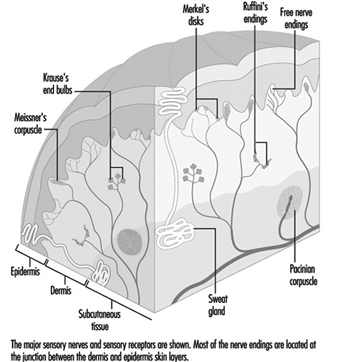
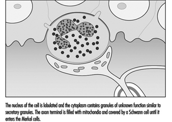
Figure 2. The touch dome on each raised region of skin contains 30 to 70 Merkel cells.

SEN80F2A

Figure 3. At a higher magnification available with the electron microscope, the Merkel cell, a specialized epithelial cell, is seen to be attached to the basement membrane that separates the epidermis from the dermis.

SEN80F2B

The Merkel cell neurite complex transduces mechanical energy into nerve impulses. While little is known about the cell’s role or about its mechanism of transduction, it has been identified as a slowly adapting receptor. This means that pressure on a touch dome containing Merkel cells causes the receptors to produce nerve impulses for the duration of the stimulus. These impulses rise in frequency in proportion to the intensity of the stimulus, thereby informing the brain of the duration and magnitude of pressure on the skin.

Like the Merkel cell, a second slowly adapting receptor also serves the skin by signalling the magnitude and duration of steady skin pressures. Visible only through a microscope, this receptor, known as the Ruffini receptor, consists of a group of neurites emerging from a myelinated fibre and encapsulated by connective tissue cells. Within the capsule structure are fibres that apparently transmit local skin distortions to the neurites, which in turn produce the messages sent along the neural highway to the central nervous system. Pressure on the skin causes a sustained discharge of nerve impulses; as with the Merkel cell, the frequency of nerve impulses is proportional to the intensity of the stimulus.

Despite their similarities, there is one outstanding difference between Merkel cells and Ruffini receptors. Whereas sensation results when Ruffini receptors are stimulated, stimulation of touch domes housing Merkel cells produces no conscious sensation; the touch dome is thus a mystery receptor, for its actual role in neural function remains unknown. Ruffini receptors, then, are believed to be the only receptors capable of providing the neural signals necessary for the sensory experience of pressure, or constant touch. In addition, it has been shown that the slowly adapting Ruffini receptors account for the ability of humans to rate cutaneous pressure on a scale of intensity.

Receptors that Respond to Vibration and Skin Movement

In contrast with slowly adapting mechanoreceptors, rapidly adapting receptors remain silent during sustained skin indentation. They are, however, well-suited to signal vibration and skin movement. Two general categories are noted: those in hairy skin, which are associated with individual hairs; and those which form corpuscular endings in glabrous, or hairless, skin.

Receptors serving hairs

A typical hair is enveloped by a network of nerve terminals branching from five to nine large myelinated axons (figure 4). In primates, these terminals fall into three categories: lanceolate endings, spindle-like terminals, and papillary endings. All three are rapidly adapting, such that a steady deflection of the hair causes nerve impulses only while movement occurs. Thus, these receptors are exquisitely sensitive to moving or vibratory stimuli, but provide little or no information about pressure, or constant touch.

Figure 4. The shafts of hairs are a platform for nerve terminals that detect movements.

SEN080F3

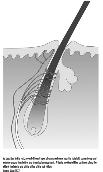
Lanceolate endings arise from a heavily myelinated fibre that forms a network around the hair. The terminal neurites lose their usual coverage of Schwann cells and work their way among the cells at the base of the hair.

Spindle-like terminals are formed by axon terminals surrounded by Schwann cells. The terminals ascend to the sloping hair shaft and end in a semicircular cluster just below a sebaceous, or oil-producing, gland. Papillary endings differ from spindle-like terminals because instead of ending on the hair shaft, they terminate as free nerve endings around the orifice of the hair.

There are, presumably, functional differences among the receptor types found on hairs. This can be inferred in part from structural differences in the way the nerves end on the hair shaft and in part from differences in the diameter of axons, as axons of different diameters connect to different central relay regions. Still, the functions of receptors in hairy skin remains an area for study.


Receptors in glabrous skin

The correlation of a receptor’s anatomical structure with the neural signals it generates is most pronounced in large and easily manipulable receptors with corpuscular, or encapsulated, endings. Particularly well understood are the pacininan and Meissner corpuscles, which, like the nerve endings in hairs discussed above, convey sensations of vibration.

The pacinian corpuscle is large enough to be seen with the naked eye, making it easy to link the receptor with a specific neural response. Located in the dermis, usually around tendons or joints, it is an onion-like structure, measuring 0.5 × 1.0 mm. It is served by one of the body’s largest afferent fibres, having a diameter of 8 to 13 μm and conducting at 50 to 80 metres per second. Its anatomy, well-studied by both light and electron microscopy, is well known.

The principal component of the corpuscle is an outer core formed of cellular material enclosing fluid-filled spaces. The outer core itself is then surrounded by a capsule that is penetrated by a central canal and a capillary network. Passing through the canal is a single myelinated nerve fibre 7 to 11 mm in diameter, which becomes a long, nonmyelinated nerve terminal that probes deep into the centre of the corpuscle. The terminal axon is elliptical, with branch-like processes.

The pacinian corpuscle is a rapidly adapting receptor. When subjected to sustained pressure, it thus produces an impulse only at the beginning and the end of the stimulus. It responds to high-frequency vibrations (80 to 400 Hz) and is most sensitive to vibrations around 250 Hz. Often, these receptors respond to vibrations transmitted along bones and tendons, and because of their extreme sensitivity, they may be activated by as little as a puff of air on the hand (Martin 1985).

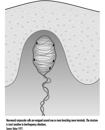
In addition to the pacinian corpuscle, there is another rapidly adapting receptor in glabrous skin. Most researchers believe it to be the Meissner corpuscle, located in the dermal papillae of the skin. Responsive to low-frequency vibrations of 2 to 40 Hz, this receptor consists of the terminal branches of a medium-sized myelinated nerve fibre enveloped in one or several layers of what appear to be modified Schwann cells, called laminar cells. The receptor’s neurites and laminar cells may connect to a basal cell in the epidermis (figure 5).

Figure 5. The Meissner corpuscle is a loosely encapsulated sensory receptor in the dermal papillae of glabrous skin.

SEN080F4

If the Meissner corpuscle is selectively inactivated by the injection of a local anaesthetic through the skin, the sense of flutter or low-frequency vibration is lost. This suggests that it functionally complements the high frequency capacity of the pacinian corpuscles. Together, these two receptors provide neural signals sufficient to account for human sensibility to a full range of vibrations (Mountcastle et al. 1967).


Cutaneous Receptors Associated with Free Nerve Endings

Many still unidentifiable myelinated and unmyelinated fibres are found in the dermis. A large number are only passing through, on their way to skin, muscles, or periosteum, while others (both myelinated and unmyelinated) appear to end in the dermis. With a few exceptions, such as the pacinian corpuscle, most fibres in the dermis appear to end in poorly defined ways or simply as free nerve endings.

While more anatomical study is needed to differentiate these ill-defined endings, physiological research has clearly shown that these fibres encode a variety of environmental events. For example, free nerve endings found at the junction between the dermis and epidermis are responsible for encoding the environmental stimuli that will be interpreted as cold, warmth, heat, pain, itch, and tickle. It is not yet known which of these different classes of small fibres convey particular sensations.

The apparent anatomical similarity of these free nerve endings is probably due to the limitations of our investigative techniques, since structural differences among free nerve endings are slowly coming to light. For example, in glabrous skin, two different terminal modes of free nerve endings have been distinguished: a thick, short pattern and a long, thin one. Studies of human hairy skin have demonstrated histochemically recognizable nerve endings that terminate at the dermal-epidermal junction: the penicillate and papillary endings. The former arise from unmyelinated fibres and form a network of endings; in contrast, the latter arise from myelinated fibres and end around the hair orifices, as mentioned earlier. Presumably, these structural disparities correspond to functional differences.

Although it is not yet possible to assign specific functions to individual structural entities, it is clear from physiological experiments that there exist functionally different categories of free nerve endings. One small myelinated fibre has been found to respond to cold in humans. Another unmyelinated fibre serving free nerve endings responds to warmth. How one class of free nerve endings can respond selectively to a drop in temperature, while an increase of skin temperature can provoke another class to signal warmth is unknown. Studies show that activation of one small fibre with a free ending may be responsible for itching or tickling sensations, while there are believed to be two classes of small fibres specifically sensitive to noxious mechanical and noxious chemical or thermal stimuli, providing the neural basis for pricking and burning pain (Keele 1964).

The definitive correlation between anatomy and physiological response awaits the development of more advanced techniques. This is one of the major stumbling blocks in the management of disorders such as causalgia, paraesthesia, and hyperpathia, which continue to present a dilemma to the physician.

Peripheral Nerve Injury

Neural function can be divided into two categories: sensory and motor. Peripheral nerve injury, usually resulting from the crushing or severing of a nerve, can impair either function or both, depending on the types of fibres in the damaged nerve. Certain aspects of motor loss tend to be misinterpreted or overlooked, as these signals do not go to muscles but rather affect autonomic vascular control, temperature regulation, the nature and thickness of the epidermis, and the condition of cutaneous mechano-receptors. The loss of motor innervation will not be discussed here, nor will the loss of innervation affecting senses other than those responsible for cutaneous sensation.

The loss of sensory innervation to the skin creates a vulnerability to further injury, as it leaves an anaesthetic surface that is incapable of signalling potentially harmful stimuli. Once injured, anaesthetized skin surfaces are slow to heal, perhaps in part on account of the lack of autonomic innervation that normally regulates such key factors as temperature regulation and cellular nutrition.

Over a period of several weeks, denervated cutaneous sensory receptors begin to atrophy, a process which is easy to observe in large encapsulated receptors such as pacinian and Meissner corpuscles. If regeneration of the axons can occur, recovery of function may follow, but the quality of the recovered function will depend upon the nature of the original injury and upon the duration of denervation (McKinnon and Dellon 1988).

Recovery following a nerve crush is more rapid, much more complete and more functional than is recovery after a nerve is severed. Two factors explain the favourable prognosis for a nerve crush. First, more axons may again achieve contact with the skin than after a transection; second, the connections are guided back to their original site by Schwann cells and linings known as basement membranes, both of which remain intact in a crushed nerve, whereas after a nerve transection the nerves often travel to incorrect regions of the skin surface by following the wrong Schwann cell paths. The latter situation results in distorted spatial information being sent to the somatosensory cortex of the brain. In both cases, however, regenerating axons appear capable of finding their way back to the same class of sensory receptors that they previously served.

The reinnervation of a cutaneous receptor is a gradual process. As the growing axon reaches the skin surface, receptive fields are smaller than normal, while the threshold is higher. These receptive points expand with time and gradually coalesce into larger fields. Sensitivity to mechanical stimuli becomes greater and often approaches the sensitivity of normal sensory receptors of that class. Studies using the stimuli of constant touch, moving touch, and vibration have shown that the sensory modalities attributed to different types of receptors return to anaesthetic areas at different rates.

Viewed under a microscope, denervated glabrous skin is seen to be thinner than normal, having flattened epidermal ridges and fewer layers of cells. This confirms that nerves have a trophic, or nutritional, influence on skin. Soon after innervation returns, the dermal ridges become better developed, the epidermis becomes thicker, and axons can be found penetrating the basement membrane. As the axon comes back to the Meissner corpuscle, the corpuscle begins to increase in size, and the previously flattened, atrophic structure returns to its original form. If the denervation has been of long duration, a new corpuscle may form adjacent to the original atrophic skeleton, which remains denervated (Dellon 1981).

As can be seen, an understanding of the consequences of peripheral nerve injury requires knowledge of normal function as well as the degrees of functional recovery. While this information is available for certain nerve cells, others require further investigation, leaving a number of murky areas in our grasp of the role of cutaneous nerves in health and disease.


Back

Additional Info

Read 23092 times Last modified on Tuesday, 11 October 2011 21:04
More in this category: « Smell

" DISCLAIMER: The ILO does not take responsibility for content presented on this web portal that is presented in any language other than English, which is the language used for the initial production and peer-review of original content. Certain statistics have not been updated since the production of the 4th edition of the Encyclopaedia (1998)."

Contents

Sensory Systems References

Adler, FH. 1992. Physiology of the Eye: Clinical Application. St. Louis: Mosby New York Books.

Adrian, WK. 1993. Visual Performance, Acuity and Age: Lux Europa Proceedings of the VIIth European Lighting Conference. London: CIBSE.

Ahlström, R, B Berglund, and U Berblund. 1986. Impaired odor perception in tank cleaners. Scand J Work Environ Health 12:574-581.

Amoore, JE. 1986. Effects of chemical exposure on olfaction in humans. In Toxicology of the Nasal Passages, edited by CS Barrow. Washington, DC: Hemisphere Publishing.

Andersen, HC, I Andersen, and J Solgard. 1977. Nasal cancers, symptoms and upper airway function in woodworkers. Br J Ind Med 34:201-207.

—. 1993. Otolaryngol Clin N Am 5(26).

Axéll, T, K Nilner, and B Nilsson. 1983. Clinical evaluation of patients referred with symptoms related to oral galvanism. Scand Dent J 7:169-178.

Ballantyne, JC and JM Ajodhia. 1984. Iatrogenic dizziness. In Vertigo, edited by MR Dix and JD Hood. Chichester: Wiley.

Bar-Sela, S, M Levy, JB Westin, R Laster, and ED Richter. 1992. Medical findings in nickel-cadmium battery workers. Israel J Med Sci 28:578-583.

Bedwal, RS, N Nair, and MP Sharma. 1993. Selenium-its biological perspectives. Med Hypoth 41:150-159.

Bell, IR. 1994. White paper: Neuropsychiatric aspects of sensitivity to low-level chemicals: A neural sensitization model. Toxicol Ind Health 10:277-312.

Besser, R, G Krämer, R Thümler, J Bohl, L Gutmann, and HC Hopf. 1987. Acute trimethyltin limbic cerebellar syndrome. Neurology 37:945-950.

Beyts, JP. 1987. Vestibular rehabilitation. In Adult Audiology, Scott-Brown’s Otolaryngology, edited by D Stephens. London: Butterworths.

Blanc, PD, HA Boushey, H Wong, SF Wintermeyer and MS Bernstein. 1993. Cytokines in metal fume fever. Am Rev Respir Dis 147:134-138.

Blount, BW. 1990. Two types of metal fume fever: mild vs. serious. Mil Med (Aug) 155(8):372-7

Bokina, AI, ND Eksler, and AD Semenenko. 1976. Investigation of the mechanism of action of atmospheric pollutants on the cenral nervous system and comparative evaluation of methods of study. Environ Health Persp 13:37-42.

Bolla, KI, BS Schwartz, and W Stewart. 1995. Comparison of neurobehavioral function in workers exposed to a mixture of organic and inorganic lead and in workers exposed to solvents. Am J Ind Med 27:231-246.

Bonnefoi, M, TM Monticello, and KT Morgan. 1991. Toxic and neoplastic responses in the nasal passages: Future research needs. Exp Lung Res 17:853-868.

Boysen, M and Solberg. 1982. Changes in the nasal mucosa of furniture workers. Scand J Work Environ Health :273-282.

Brittebo, EB, PG Hogman, and I Brandt. 1987. Epithelial binding of hexachlorocyclohexanes in the respiratory and upper alimentary tracts: A comparison between the alpha-, beta-, and gamma-isomers in mice. Food Chem Toxicol 25:773-780.

Brooks, SM. 1994. Host susceptibility to indoor air pollution. J Allergy Clin Immunol 94:344-351.

Callender, TJ, L Morrow, K Subramanian, D Duhon, and M Ristovv. 1993. Three-dimensional brain metabolic imaging in patients with toxic encephalopathy. Environmental Research 60:295-319.

Chia, SE, CN Ong, SC Foo, and HP Lee. 1992. Medical student’s exposure to formaldehyde in a gross anatomy dissection laboratory. J Am Coll Health 41:115-119.

Choudhuri, S, KK Kramer, and NE Berman. 1995. Constitutive expression of metallothionein genes in mouse brain. Toxicol Appl Pharmacol 131:144-154.

Ciesielski, S, DP Loomis, SR Mims, and A Auer. 1994. Pesticide exposures, cholinesterase depression, and symptoms among North Carolina migrant farmworkers. Am J Public Health 84:446-451.

Clerisi, WJ, B Ross, and LD Fechter. 1991. Acute ototoxicity of trialkyltins in the guinea pig. Toxicol Appl Pharmacol :547-566.

Coleman, JW, MR Holliday, and RJ Dearman. 1994. Cytokine-mast cell interactions: Relevance to IgE-mediated chemical allergy. Toxicology 88:225-235.

Cometto-Muñiz, JE and WS Cain. 1991. Influence of airborne contaminants on olfaction and the common chemical sense. In Smell and Taste in Health and Disease, edited by TV Getchell. New York: Raven Press.

—. 1994. Sensory reactions of nasal pungency and odor to volatile organic compounds: The alkylbenzenes. Am Ind Hyg Assoc J 55:811-817.

Corwin, J, M Loury, and AN Gilbert. 1995. Workplace, age, and sex as mediators of olfactory function: Data from the National Geographic Smell Survey. Journal of Gerontolgy: Psychiol Sci 50B:P179-P186.

Council on Dental Materials, Instruments and Equipment. 1987. American Dental Association status report on the occurence of galvanic corrosion in the mouth and its potential effects. J Am Dental Assoc 115:783-787.

Council on Scientific Affairs. 1989. Council report: Formaldehyde. JAMA 261:1183-1187.

Crampton, GH. 1990. Motion and Space Sickness. Boca Raton: CRC Press.

Cullen, MR. 1987. Workers with multiple chemical sensitivities. Occup Med: State Art Rev 2(4).

Deems, DA, RL Doty, and RG Settle. 1991. Smell and taste disorders, a study of 750 patients from the University of Pennsylvania Smell and Taste Center. Arch Otolaryngol Head Neck Surg 117:519-528.

Della Fera, MA, AE Mott, and ME Frank. 1995. Iatrogenic causes of taste disturbances: Radiation therapy, surgery, and medication. In Handbook of Olfaction and Gustation, edited by RL Doty. New York: Marcel Dekker.

Dellon, AL. 1981. Evaluation of Sensibility and Re-Education of Sensation in the Hand. Baltimore: Williams & Wilkins.

Dykes, RW. 1977. Sensory receptors. In Reconstructive Microsurgery, edited by RK Daniel and JK Terzis. Boston: Little Brown & Co.

El-Etri, MM, WT Nickell, M Ennis, KA Skau, and MT Shipley. 1992. Brain norepinephrine reductions in soman-intoxicated rats: Association with convulsions and AchE inhibition, time course, and relation to other monoamines. Experimental Neurology 118:153-163.

Evans, J and L Hastings. 1992. Accumulation of Cd(II) in the CNS depending on the route of administration: Intraperitoneal, intratracheal, or intranasal. Fund Appl Toxicol 19:275-278.

Evans, JE, ML Miller, A Andringa, and L Hastings. 1995. Behavioral, histological, and neurochemical effets of nickel(II) on the rat olfactory system. Toxicol Appl Pharmacol 130:209-220.

Fechter, LD, JS Young, and L Carlisle. 1988. Potentiation of noise induced threshold shifts and hair cell loss by carbon monoxide. Hearing Res 34:39-48.
Fox, SL. 1973. Industrial and Occupational Opthalmology. Springfield: Charles C. Thomas.

Frank, ME, TP Hettinger, and AE Mott. 1992. The sense of taste: Neurobiology, aging, and medication effects. Critical Reviews in Oral Biology Medicine 3:371-393.

Frank, ME and DV Smith. 1991. Electrogustometry: A simple way to test taste. In Smell and Taste in Health and Disease, edited by TV Getchell, RL Doty, and LM Bartoshuk. New York: Raven Press.

Gagnon, P, D Mergler, and S Lapare. 1994. Olfactory adaptation, threshold shift and recovery at low levels of exposure to methyl isobutyl ketone (MIBK). Neurotoxicology 15:637-642.

Gilbertson, TA. 1993. The physiology of vertebrate taste reception. Curr Opin Neurobiol 3:532-539.

Gordon, T and JM Fine. 1993. Metal fume fever. Occup Med: State Art Rev 8:505-517.

Gosselin, RE, RP Smith, and HC Hodge. 1984. Clinical Toxicology of Commercial Products. Baltimore: Williams & Wilkins.

Graham, CH, NR Barlett, JL Brown, Y Hsia, CG Mueller, and LA Riggs. 1965. Vision and Visual Perception. New York: John Wiley and Sons, Inc.

Grandjean, E. 1987. Ergonomics in Computerized Offices. London: Taylor & Francis.

Grant, A. 1979. Optical danger of fiberglass hardener. Med J Austral 1:23.

Gresham, LS, CA Molgaard, and RA Smith. 1993. Induction of cytochrome P-450 enzymes via tobacco smoke: A potential mechanism for developing resistance to environmental toxins as related to Parkinsonism and other neurologic disease. Neuroepidemiol 12:114-116.

Guidotti, TL. 1994. Occupational exposure to hydrogen sulfide in the sour gas industry: Some unresolved issues. Int Arch Occup Environ Health 66:153-160.

Gyntelberg, F, S Vesterhauge, P Fog, H Isager, and K Zillstorff. 1986. Acquired intolerance to organic solvents and results of vestibular testing. Am J Ind Med 9:363-370.

Hastings, L. 1990. Sensory neurotoxicology: use of the olfactory system in the assessment of toxicity. Neurotoxicology and Teratology 12:455-459.

Head, PW. 1984. Vertigo and barotrauma. In Vertigo, edited by MR Dix and JD Hood. Chichester: Wiley.

Hohmann, B and F Schmuckli. 1989. Dangers du bruit pour l’ouië et l’emplacement de travail. Lucerne: CNA.

Holmström, M, G Rosén, and B Wilhelmsson. 1991. Symptoms, airway physiology and histology of workers exposed to medium-density fiber board. Scand J Work Environ Health 17:409-413.

Hotz, P, A Tschopp, D Söderström, and J Holtz. 1992. Smell or taste disturbances, neurological symptoms, and hydrocarbon exposure. Int Arch Occup Environ Health 63:525-530.

Howard, IP. 1982. Human Visual Orientation. Chichester: Wiley.

Iggo, A and AR Muir. 1969. The structure and function of a slowly adapting touch corpuscle in hairy skin. J Physiol Lond 200(3):763-796.

Illuminating Engineering Society of North America (IESNA). 1993. Vision and perception. In Lighting Handbook: Reference and Application, edited by MS Rea and Fies. New York: IESNA.

Innocenti, A, M Valiani, G Vessio, M Tassini, M Gianelli, and S Fusi. 1985. Wood dust and nasal diseases: Exposure to chestnut wood dust and loss of smell (pilot study). Med Lavoro 4:317-320.

Jacobsen, P, HO Hein, P Suadicani, A Parving, and F Gyntelberg. 1993. Mixed solvent exposure and hearing impairment: An epidemiological study of 3284 men. The Copenhagen male study. Occup Med 43:180-184.

Johansson, B, E Stenman, and M Bergman. 1984. Clinical study of patients referred for investigation regarding so-called oral galvanism. Scand J Dent Res 92:469-475.

Johnson, A-C and PR Nylén. 1995. Effects of industrial solvents on hearing. Occup Med: State of the art reviews. 10:623-640.

Kachru, DM, SK Tandon, UK Misra, and D Nag. 1989. Occupational lead poisoning among silver jewelry workers. Indian Journal of Medical Sciences 43:89-91.

Keele, CA. 1964. Substances Producing Pain and Itch. London: Edward Arnold.

Kinnamon, SC and TV Getchell. 1991. Sensory transduction in olfactory receptor neurons and gustatory receptor cells. In Smell and Taste in Health and Disease, edited by TV Getchell, RL Doty, and LM Bartoshuk. New York: Raven Press.

Krueger, H. 1992. Exigences visuelles au poste de travail: Diagnostic et traitement. Cahiers
médico-sociaux 36:171-181.

Lakshmana, MK, T Desiraju, and TR Raju. 1993. Mercuric chloride-induced alterations of levels of noradrenaline, dopamine, serotonin and acetylcholine esterase activity in different regions of rat brain during postnatal development. Arch Toxicol 67:422-427.

Lima, C and JP Vital. 1994. Olfactory mucosa response in guinea pigs following intranasal instillation with Cryptococcus neoformans: A histological and immunocytochemical study. Mycopathologia 126:65-73.

Luxon, LM. 1984. The anatomy and physiology of the vestibular system. In Vertigo, edited by MR Dix and JD Hood. Chichester: Wiley.

MacKinnon, SE and AL Dellon. 1988. Surgery of the Peripheral Nerve. New York: Thieme Medical Publishers.

Marek, J-J. 1993. The molecular biology of taste transduction. Bioessays 15:645-650.

Marek, M. 1992. Interactions between dental amalgams and the oral environment. Adv Dental Res 6:100-109.

Margolskee, RF. 1993. The biochemistry and molecular biology of taste transduction. Curr Opin Neurobiol 3:526-531.

Martin, JH. 1985. Receptor physiology and submodality coding in the somatic sensory system. Principles of Neuroscience, edited by ER Kandel and JH Schwartz.

Meyer, J-J. 1990. Physiologie de la vision et ambiance lumineuse. Document de l’Aerospatiale, Paris.

Meyer, J-J, A Bousquet, L Zoganas and JC Schira. 1990. Discomfort and disability glare in VDT operators. In Work with Display Units 89, edited by L Berlinguet and D Berthelette. Amsterdam: Elsevier Science.

Meyer, J-J, P Rey, and A Bousquet. 1983. An automatic intermittent light stimulator to record flicker perceptive thresholds in patients with retinal disease. In Advances in Diagnostic Visual Optics, edited by GM Brenin and IM Siegel. Berlin: Springer-Verlag.

Meyer, J-J, P Rey, B Thorens, and A Beaumanoire. 1971. Examen de sujets atteints d’un traummatisme cranio-cérébral par un test perception visuelle: courbe de Lange. Swiss Arch of Neurol 108:213-221.

Meyer, J-J, A Bousquet, JC Schira, L Zoganas, and P Rey. 1986. Light sensitivity and visual strain when driving at night. In Vision in Vehicles, edited by AG Gale. Amsterdam: Elsevier Science Publisher.

Miller, CS. 1992. Possible models for multiple chemical sensitivity: conceptual issues and role of the limbic system. Toxicol Ind Health 8:181-202.

Miller, RR, JT Young, RJ Kociba, DG Keyes, KM Bodner, LL Calhoun, and JA Ayres. 1985. Chronic toxicity and oncogenicity bioassay of inhaled ethyl acrylate in fischer 344 rats and B6C3F1 mice. Drug Chem Toxicol 8:1-42.

Möller, C, L Ödkvist, B Larsby, R Tham, T Ledin, and L Bergholtz. 1990. Otoneurological finding among workers exposed to styrene. Scand J Work Environ Health 16:189-194.

Monteagudo, FSE, MJD Cassidy, and PI Folb. 1989. Recent developments in aluminum toxicology. Med Toxicol 4:1-16.

Morata, TC, DE Dunn, LW Kretschmer, GK Lemasters, and RW Keith. 1993. Effects of occupational exposure to organic solvents and noise on hearing. Scand J Work Environ Health 19:245-254.

Mott, AE, M Grushka, and BJ Sessle. 1993. Diagnosis and management of taste disorders and burning mouth syndrome. Dental Clinics of North America 37:33-71.

Mott, AE and DA Leopold. 1991. Disorders in taste and smell. Med Clin N Am 75:1321-1353.

Mountcastle, VB. 1974. Medical Physiology. St. Louis: CV Mosby.

Mountcastle, VB, WH Talbot, I Darian-Smith, and HH Kornhuber. 1967. Neural basis of the sense of flutter-vibration. Science :597-600.

Muijser, H, EMG Hoogendijk, and J Hoosima. 1988. The effects of occupational exposure to styrene on high-frequency hearing thresholds. Toxicology :331-340.

Nemery, B. 1990. Metal toxicity and the respiratory tract. Eur Respir J 3:202-219.

Naus, A. 1982. Alterations of the smell acuity caused by menthol. J Laryngol Otol 82:1009-1011.

Örtendahl, TW. 1987. Oral changes in divers working with electrical welding/cutting underwater. Swedish Dent J Suppl 43:1-53.

Örtendahl, TW, G Dahlén, and HOE Röckert. 1985. The evaluation of oral problems in divers performing electrical welding and cutting under water. Undersea Biomed Res 12:55-62.

Ogawa, H. 1994. Gustatory cortex of primates: Anatomy and physiology. Neurosci Res 20:1-13.

O’Reilly, JP, BL Respicio, and FK Kurata. 1977. Hana Kai II: A 17-day dry saturation dive at 18.6 ATA. VII: Auditory, visual and gustatory sensations. Undersea Biomed Res 4:307-314.

Otto, D, G Robinson, S Bauman, S Schroeder, P Mushak, D Kleinbaum, and L Boone. 1985. %-years follow-up study of children with low-to-moderate lead absorption: Electrophysiological evaluation. Environ Research 38:168-186.

Oyanagi, K, E Ohama, and F Ikuta. 1989. The auditory system in methyl mercurial intoxication: A neuropathological investigation on 14 autopsy cases in Niigata, Japan. Acta Neuropathol 77:561-568.

Participants of SCP Nos. 147/242 and HF Morris. 1990. Veterans administration cooperative studies project no. 147: Association of metallic taste with metal ceramic alloys. J Prosthet Dent 63:124-129.

Petersen, PE and C Gormsen. 1991. Oral conditions among German battery factory workers. Community Dentistry and Oral Epidemiology 19:104-106.

Pfeiffer, P and H Schwickerath. 1991. Nickel solubility and metallic taste. Zwr 100:762-764,766,768-779.

Pompeiano, O and JHJ Allum. 1988. Vestibulospinal Control of Posture and Locomotion. Progress in Brain Research, no.76. Amsterdam: Elsevier.

Rees, T and L Duckert. 1994. Hearing loss and other otic disorders. In Textbook of Clinical, Occupational and Environmental Medicine, edited by C Rosenstock. Philadelphia: WB Saunders.

Ressler, KJ, SL Sullivan, and LB Buck. 1994. A molecular dissection of spatial patterning in the olfactory system. Curr Opin Neurobiol 4:588-596.

Rey, P. 1991. Précis De Medecine Du Travail. Geneva: Medicine et Hygiène.

Rey, P and A Bousquet. 1990. Medical eye examination strategies for VDT operators. In Work With Display Units 89, edited by L Berlinguet and D Berthelette. Amsterdam: Elsevier Science.

Rose, CS, PG Heywood, and RM Costanzo. 1934. Olfactory impairment after chronic occupational cadmium exposure. J Occup Med 34:600-605.

Rubino, GF. 1990. Epidemiologic survey of ocular disorders: The Italian multicentric research. In Work with Display Units 89, edited by L Berlinguet and D Berthelette. Amsterdam: Elsevier Science Publishers B.V.

Ruth, JH. 1986. Odor thresholds and irritation levels of several chemical substances: A review. Am Ind Hyg Assoc J 47:142-151.

Rusznak, C, JL Devalia, and RJ Davies. 1994. The impact of pollution on allergic disease. Allergy 49:21-27.

Ryback, LP. 1992. Hearing: The effects of chemicals. Otolaryngology-Head and Neck Surgery 106:677-686.

—. 1993. Ototoxicity. Otolaryngol Clin N Am 5(26).

Savov, A. 1991. Damages to the ears, nose and throat in copper production. Problemi na Khigienata 16:149-153.

—. 1994. Changes in taste and smell: Drug interactions and food preferences. Nutr Rev 52(II):S11-S14.

Schiffman, SS. 1994. Changes in taste and smell: Drug interactions and food preferences. Nutr Rev 52(II): S11-S14.

Schiffman, SS and HT Nagle. 1992. Effect of environmental pollutants on taste and smell. Otolaryngology-Head and Neck Surgery 106:693-700.

Schwartz, BS, DP Ford, KI Bolla, J Agnew, and ML Bleecker. 1991. Solvent-associated olfatory dysfunction: Not a predictor of deficits in learning and memory. Am J Psychiatr 148:751-756.

Schweisfurth, H and C Schottes. 1993. Acute intoxication of a hydrazine-like gas by 19 workers in a garbage dump. Zbl Hyg 195:46-54.

Shusterman, D. 1992. Critical review: The health significance of environmental odor pollution. Arch Environ Health 47:76-87.

Shusterman, DJ and JE Sheedy. 1992. Occupational and environmental disorders of the special senses. Occup Med: State Art Rev 7:515-542.

Siblerud, RL. 1990. The relationship between mercury from dental amalgam and oral cavity health. Ann Dent 49:6-10.

Sinclair. 1981. Mechanisms of Cutaneous Sensation. Oxford: Oxford Univ. Press.

Spielman, AI. 1990. Interaction of saliva and taste. J Dental Res 69:838.

Stevens, JC and WS Cain. 1986. Aging and the perception of nasal irritation. Physiol Behav 37:323-328.

van Dijk, FJH. 1986. Non-auditory effects of noise in industry. II A review of the literature. Int Arch Occup Environ Health 58.

Verriest, G and G Hermans. 1975. Les aptitudes visuelles professionnelles. Bruxelles: Imprimerie médicale et scientifique.

Welch, AR, JP Birchall, and FW Stafford. 1995. Occupational rhinitis - Possible mechanisms of pathogenesis. J Laryngol Otol 109:104-107.

Weymouth, FW. 1966. The eye as an optical instrument. In Physiology and Biophysics, edited by TC Ruch and HD Patton. London: Saunders.

Wieslander, G, D Norbäck, and C Edling. 1994. Occupational exposure to water based paint and symptoms from the skin and eyes. Occup Environ Med 51:181-186.

Winberg, S, R Bjerselius, E Baatrup, and KB Doving. 1992. The effect of Cu(II) on the electro-olfactogram (EOG) of the Atlantic salmon (Salmo salar L) in artificial freshwater of varying inorganic carbon concentrations. Ecotoxicology and Environmental Safety 24:167-178.

Witek, TJ. 1993. The nose as a target for adverse effects from the environment: Applying advances in nasal physiologic measurements and mechanisms. Am J Ind Med 24:649-657.

World Health Organization (WHO). 1981. Arsenic. Environmental Health Criteria, no.18. Geneva: WHO.

Yardley, L. 1994. Vertigo and Dizziness. London: Routledge.

Yontchev, E, GE Carlsson, and B Hedegård. 1987. Clinical findings in patients with orofacial discomfort complaints. Int J Oral Maxillofac Surg 16:36-44.