Sunday, 16 January 2011 16:29

Cellular Injury and Cellular Death

Rate this item
(1 Vote)

Virtually all of medicine is devoted to either preventing cell death, in diseases such as myocardial infarction, stroke, trauma and shock, or causing it, as in the case of infectious diseases and cancer. It is, therefore, essential to understand the nature and mechanisms involved. Cell death has been classified as “accidental”, that is, caused by toxic agents, ischaemia and so on, or “programmed”, as occurs during embryological development, including formation of digits, and resorption of the tadpole tail.

Cell injury and cell death are, therefore, important both in physiology and in pathophysiology. Physiological cell death is extremely important during embryogenesis and embryonic development. The study of cell death during development has led to important and new information on the molecular genetics involved, especially through the study of development in invertebrate animals. In these animals, the precise location and the significance of cells that are destined to undergo cell death have been carefully studied and, with the use of classic mutagenesis techniques, several involved genes have now been identified. In adult organs, the balance between cell death and cell proliferation controls organ size. In some organs, such as the skin and the intestine, there is a continual turnover of cells. In the skin, for example, cells differentiate as they reach the surface, and finally undergo terminal differentiation and cell death as keratinization proceeds with the formation of crosslinked envelopes.

Many classes of toxic chemicals are capable of inducing acute cell injury followed by death. These include anoxia and ischaemia and their chemical analogues such as potassium cyanide; chemical carcinogens, which form electrophiles that covalently bind to proteins in nucleic acids; oxidant chemicals, resulting in free radical formation and oxidant injury; activation of complement; and a variety of calcium ionophores. Cell death is also an important component of chemical carcinogenesis; many complete chemical carcinogens, at carcinogenic doses, produce acute necrosis and inflammation followed by regeneration and preneoplasia.

Definitions

Cell injury

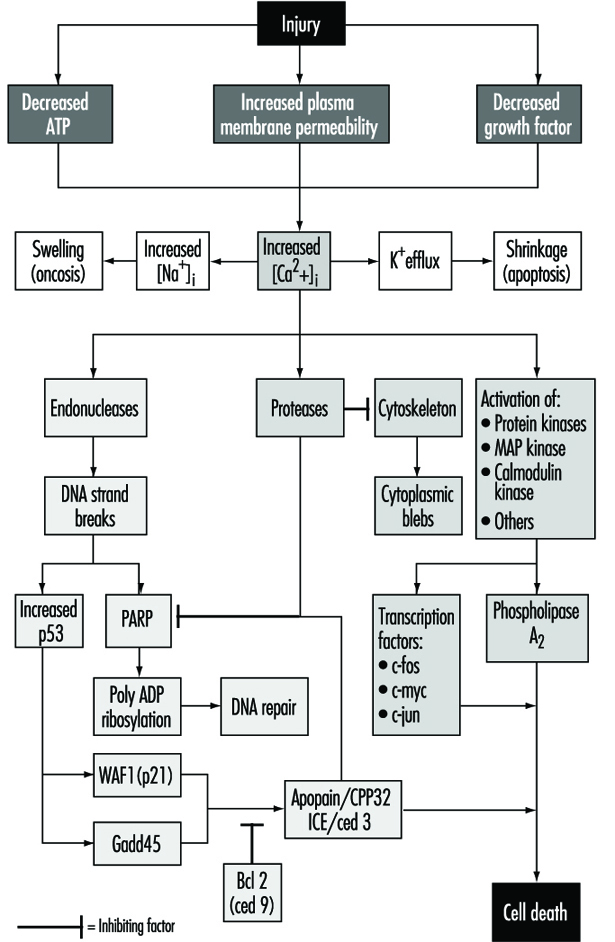
Cell injury is defined as an event or stimulus, such as a toxic chemical, that perturbs the normal homeostasis of the cell, thus causing a number of events to occur (figure 1). The principal targets of lethal injury illustrated are inhibition of ATP synthesis, disruption of plasma membrane integrity or withdrawal of essential growth factors.

Figure 1. Cell injury

TOX060F1

Lethal injuries result in the death of a cell after a variable period of time, depending on temperature, cell type and the stimulus; or they can be sublethal or chronic—that is, the injury results in an altered homeostatic state which, though abnormal, does not result in cell death (Trump and Arstila 1971; Trump and Berezesky 1992; Trump and Berezesky 1995; Trump, Berezesky and Osornio-Vargas 1981). In the case of a lethal injury, there is a phase prior to the time of cell death

during this time, the cell will recover; however, after a particular point in time (the “point of no return” or point of cell death), the removal of the injury does not result in recovery but instead the cell undergoes degradation and hydrolysis, ultimately reaching physical-chemical equilibrium with the environment. This is the phase known as necrosis. During the prelethal phase, several principal types of change occur, depending on the cell and the type of injury. These are known as apoptosis and oncosis.


Apoptosis

Apoptosis is derived from the Greek words apo, meaning away from, and ptosis, meaning to fall. The term falling away from is derived from the fact that, during this type of prelethal change, the cells shrink and undergo marked blebbing at the periphery. The blebs then detach and float away. Apoptosis occurs in a variety of cell types following various types of toxic injury (Wyllie, Kerr and Currie 1980). It is especially prominent in lymphocytes, where it is the predominant mechanism for turnover of lymphocyte clones. The resulting fragments result in the basophilic bodies seen within macrophages in lymph nodes. In other organs, apoptosis typically occurs in single cells which are rapidly cleared away before and following death by phagocytosis of the fragments by adjacent parenchymal cells or by macrophages. Apoptosis occurring in single cells with subsequent phagocytosis typically does not result in inflammation. Prior to death, apoptotic cells show a very dense cytosol with normal or condensed mitochondria. The endoplasmic reticulum (ER) is normal or only slightly dilated. The nuclear chromatin is markedly clumped along the nuclear envelope and around the nucleolus. The nuclear contour is also irregular and nuclear fragmentation occurs. The chromatin conden- sation is associated with DNA fragmentation which, in many instances, occurs between nucleosomes, giving a characteristic ladder appearance on electrophoresis.

In apoptosis, increased [Ca2+]i may stimulate K+ efflux resulting in cell shrinkage, which probably requires ATP. Injuries that totally inhibit ATP synthesis, therefore, are more likely to result in apoptosis. A sustained increase of [Ca2+]i has a number of deleterious effects including activation of proteases, endonucleases, and phospholipases. Endonuclease activation results in single and double DNA strand breaks which, in turn, stimulate increased levels of p53 and in poly-ADP ribosylation, and of nuclear proteins which are essential in DNA repair. Activation of proteases modifies a number of substrates including actin and related proteins leading to bleb formation. Another important substrate is poly(ADP-ribose) polymerase (PARP), which inhibits DNA repair. Increased [Ca2+]i is also associated with activation of a number of protein kinases, such as MAP kinase, calmodulin kinase and others. Such kinases are involved in activation of transcription factors which initiate transcription of immediate-early genes, for example, c-fos, c-jun and c-myc, and in activation of phospholipase A2 which results in permeabilization of the plasma membrane and of intracellular membranes such as the inner membrane of mitochondria.

Oncosis

Oncosis, derived from the Greek word onkos, to swell, is so named because in this type of prelethal change the cell begins to swell almost immediately following the injury (Majno and Joris 1995). The reason for the swelling is an increase in cations in the water within the cell. The principal cation responsible is sodium, which is normally regulated to maintain cell volume. However, in the absence of ATP or if Na-ATPase of the plasmalemma is inhibited, volume control is lost because of intracellular protein, and sodium in the water continuing to increase. Among the early events in oncosis are, therefore, increased [Na+]i which leads to cellular swelling and increased [Ca2+]i resulting either from influx from the extracellular space or release from intracellular stores. This results in swelling of the cytosol, swelling of the endoplasmic reticulum and Golgi apparatus, and the formation of watery blebs around the cell surface. The mitochondria initially undergo condensation, but later they too show high-amplitude swelling because of damage to the inner mitochondrial membrane. In this type of prelethal change, the chromatin undergoes condensation and ultimately degradation; however, the characteristic ladder pattern of apoptosis is not seen.

Necrosis

Necrosis refers to the series of changes that occur following cell death when the cell is converted to debris which is typically removed by the inflammatory response. Two types can be distinguished: oncotic necrosis and apoptotic necrosis. Oncotic necrosis typically occurs in large zones, for example, in a myocardial infarct or regionally in an organ after chemical toxicity, such as the renal proximal tubule following administration of HgCl2. Broad zones of an organ are involved and the necrotic cells rapidly incite an inflammatory reaction, first acute and then chronic. In the event that the organism survives, in many organs necrosis is followed by clearing away of the dead cells and regeneration, for example, in the liver or kidney following chemical toxicity. In contrast, apoptotic necrosis typically occurs on a single cell basis and the necrotic debris is formed within the phagocytes of macrophages or adjacent parenchymal cells. The earliest characteristics of necrotic cells include interruptions in plasma membrane continuity and the appearance of flocculent densities, representing denatured proteins within the mitochondrial matrix. In some forms of injury that do not initially interfere with mitochondrial calcium accumulation, calcium phosphate deposits can be seen within the mitochondria. Other membrane systems are similarly fragmenting, such as the ER, the lysosomes and the Golgi apparatus. Ultimately, the nuclear chromatin undergoes lysis, resulting from attack by lysosomal hydrolases. Following cell death, lysosomal hydrolases play an important part in clearing away debris with cathepsins, nucleolases and lipases since these have an acid pH optimum and can survive the low pH of necrotic cells while other cellular enzymes are denatured and inactivated.

Mechanisms

Initial stimulus

In the case of lethal injuries, the most common initial interactions resulting in injury leading to cell death are interference with energy metabolism, such as anoxia, ischaemia or inhibitors of respiration, and glycolysis such as potassium cyanide, carbon monoxide, iodo-acetate, and so on. As mentioned above, high doses of compounds that inhibit energy metabolism typically result in oncosis. The other common type of initial injury resulting in acute cell death is modification of the function of the plasma membrane (Trump and Arstila 1971; Trump, Berezesky and Osornio-Vargas 1981). This can either be direct damage and permeabilization, as in the case of trauma or activation of the C5b-C9 complex of complement, mechanical damage to the cell membrane or inhibition of the sodium-potassium (Na+-K+) pump with glycosides such as ouabain. Calcium ionophores such as ionomycin or A23187, which rapidly carry [Ca2+] down the gradient into the cell, also cause acute lethal injury. In some cases, the pattern in the prelethal change is apoptosis; in others, it is oncosis.

Signalling pathways

With many types of injury, mitochondrial respiration and oxidative phosphorylation are rapidly affected. In some cells, this stimulates anaerobic glycolysis, which is capable of maintaining ATP, but with many injuries this is inhibited. The lack of ATP results in failure to energize a number of important homeostatic processes, in particular, control of intracellular ion homeostasis (Trump and Berezesky 1992; Trump, Berezesky and Osornio-Vargas 1981). This results in rapid increases of [Ca2+]i, and increased [Na+] and [Cl-] results in cell swelling. Increases in [Ca2+]i result in the activation of a number of other signalling mechanisms discussed below, including a series of kinases, which can result in increased immediate early gene transcription. Increased [Ca2+]i also modifies cytoskeletal function, in part resulting in bleb formation and in the activation of endonucleases, proteases and phospholipases. These seem to trigger many of the important effects discussed above, such as membrane damage through protease and lipase activation, direct degradation of DNA from endonuclease activation, and activation of kinases such as MAP kinase and calmodulin kinase, which act as transcription factors.

Through extensive work on development in the invertebrate C. elegans and Drosophila, as well as human and animal cells, a series of pro-death genes have been identified. Some of these invertebrate genes have been found to have mammalian counterparts. For example, the ced-3 gene, which is essential for programmed cell death in C. elegans, has protease activity and a strong homology with the mammalian interleukin converting enzyme (ICE). A closely related gene called apopain or prICE has recently been identified with even closer homology (Nicholson et al. 1995). In Drosophila, the reaper gene seems to be involved in a signal that leads to programmed cell death. Other pro-death genes include the Fas membrane protein and the important tumour-suppressor gene, p53, which is widely conserved. p53 is induced at the protein level following DNA damage and when phosphorylated acts as a transcription factor for other genes such as gadd45 and waf-1, which are involved in cell death signalling. Other immediate early genes such as c-fos, c-jun, and c-myc also seem to be involved in some systems.

At the same time, there are anti-death genes which appear to counteract the pro-death genes. The first of these to be identified was ced-9 from C. elegans, which is homologous to bcl-2 in humans. These genes act in an as yet unknown way to prevent cell killing by either genetic or chemical toxins. Some recent evidence indicates that bcl-2 may act as an antioxidant. Currently, there is much effort underway to develop an understanding of the genes involved and to develop ways to activate or inhibit these genes, depending on the situation.


Back

Read 12054 times Last modified on Tuesday, 26 July 2022 19:28

" DISCLAIMER: The ILO does not take responsibility for content presented on this web portal that is presented in any language other than English, which is the language used for the initial production and peer-review of original content. Certain statistics have not been updated since the production of the 4th edition of the Encyclopaedia (1998)."

Contents

Toxicology References

Andersen, KE and HI Maibach. 1985. Contact allergy predictive tests on guinea pigs. Chap. 14 in Current Problems in Dermatology. Basel: Karger.

Ashby, J and RW Tennant. 1991. Definitive relationships among chemical structure, carcinogenicity and mutagenicity for 301 chemicals tested by the US NTP. Mutat Res 257:229-306.

Barlow, S and F Sullivan. 1982. Reproductive Hazards of Industrial Chemicals. London: Academic Press.

Barrett, JC. 1993a. Mechanisms of action of known human carcinogens. In Mechanisms of Carcinogenesis in Risk Identification, edited by H Vainio, PN Magee, DB McGregor, and AJ McMichael. Lyon: International Agency for Research on Cancer (IARC).

—. 1993b. Mechanisms of multistep carcinogenesis and carcinogen risk assessment. Environ Health Persp 100:9-20.

Bernstein, ME. 1984. Agents affecting the male reproductive system: Effects of structure on activity. Drug Metab Rev 15:941-996.

Beutler, E. 1992. The molecular biology of G6PD variants and other red cell defects. Annu Rev Med 43:47-59.

Bloom, AD. 1981. Guidelines for Reproductive Studies in Exposed Human Populations. White Plains, New York: March of Dimes Foundation.

Borghoff, S, B Short and J Swenberg. 1990. Biochemical mechanisms and pathobiology of a-2-globulin nephropathy. Annu Rev Pharmacol Toxicol 30:349.

Burchell, B, DW Nebert, DR Nelson, KW Bock, T Iyanagi, PLM Jansen, D Lancet, GJ Mulder, JR Chowdhury, G Siest, TR Tephly, and PI Mackenzie. 1991. The UPD-glucuronosyltransferase gene superfamily: Suggested nomenclature based on evolutionary divergence. DNA Cell Biol 10:487-494.

Burleson, G, A Munson, and J Dean. 1995. Modern Methods in Immunotoxicology. New York: Wiley.

Capecchi, M. 1994. Targeted gene replacement. Sci Am 270:52-59.

Carney, EW. 1994. An integrated perspective on the developmental toxicity of ethylene glycol. Rep Toxicol 8:99-113.

Dean, JH, MI Luster, AE Munson, and I Kimber. 1994. Immunotoxicology and Immunopharmacology. New York: Raven Press.

Descotes, J. 1986. Immunotoxicology of Drugs and Chemicals. Amsterdam: Elsevier.

Devary, Y, C Rosette, JA DiDonato, and M Karin. 1993. NFkB activation by ultraviolet light not dependent on a nuclear signal. Science 261:1442-1445.

Dixon, RL. 1985. Reproductive Toxicology. New York: Raven Press.

Duffus, JH. 1993. Glossary for chemists of terms used in toxicology. Pure Appl Chem 65:2003-2122.

Elsenhans, B, K Schuemann, and W Forth. 1991. Toxic metals: Interactions with essential metals. In Nutrition, Toxicity and Cancer, edited by IR Rowland. Boca-Raton: CRC Press.

Environmental Protection Agency (EPA). 1992. Guidelines for exposure assessment. Federal Reg 57:22888-22938.

—. 1993. Principles of neurotoxicity risk assessment. Federal Reg 58:41556-41598.

—. 1994. Guidelines for Reproductive Toxicity Assessment. Washington, DC: US EPA: Office of Research and Development.

Fergusson, JE. 1990. The Heavy Elements. Chap. 15 in Chemistry, Environmental Impact and Health Effects. Oxford: Pergamon.

Gehring, PJ, PG Watanabe, and GE Blau. 1976. Pharmacokinetic studies in evaluation of the toxicological and environmental hazard of chemicals. New Concepts Saf Eval 1(Part 1, Chapter 8):195-270.

Goldstein, JA and SMF de Morais. 1994. Biochemistry and molecular biology of the human CYP2C subfamily. Pharmacogenetics 4:285-299.

Gonzalez, FJ. 1992. Human cytochromes P450: Problems and prospects. Trends Pharmacol Sci 13:346-352.

Gonzalez, FJ, CL Crespi, and HV Gelboin. 1991. cDNA-expressed human cytochrome P450: A new age in molecular toxicology and human risk assessment. Mutat Res 247:113-127.

Gonzalez, FJ and DW Nebert. 1990. Evolution of the P450 gene superfamily: Animal-plant “warfare,” molecular drive, and human genetic differences in drug oxidation. Trends Genet 6:182-186.

Grant, DM. 1993. Molecular genetics of the N-acetyltransferases. Pharmacogenetics 3:45-50.

Gray, LE, J Ostby, R Sigmon, J Ferrel, R Linder, R Cooper, J Goldman, and J Laskey. 1988. The development of a protocol to assess reproductive effects of toxicants in the rat. Rep Toxicol 2:281-287.

Guengerich, FP. 1989. Polymorphism of cytochrome P450 in humans. Trends Pharmacol Sci 10:107-109.

—. 1993. Cytochrome P450 enzymes. Am Sci 81:440-447.

Hansch, C and A Leo. 1979. Substituent Constants for Correlation Analysis in Chemistry and Biology. New York: Wiley.

Hansch, C and L Zhang. 1993. Quantitative structure-activity relationships of cytochrome P450. Drug Metab Rev 25:1-48.

Hayes AW. 1988. Principles and Methods of Toxicology. 2nd ed. New York: Raven Press.

Heindell, JJ and RE Chapin. 1993. Methods in Toxicology: Male and Female Reproductive Toxicology. Vol. 1 and 2. San Diego, Calif.: Academic Press.

International Agency for Research on Cancer (IARC). 1992. Solar and ultraviolet radiation. Lyon: IARC.

—. 1993. Occupational Exposures of Hairdressers and Barbers and Personal Use of Hair Colourants: Some Hair Dyes, Cosmetic Colourants, Industrial Dyestuffs and Aromatic Amines. Lyon: IARC.

—. 1994a. Preamble. Lyon: IARC.

—. 1994b. Some Industrial Chemicals. Lyon: IARC.

International Commission on Radiological Protection (ICRP). 1965. Principles of Environmental Monitoring Related to the Handling of Radioactive Materials. Report of Committee IV of The International Commission On Radiological Protection. Oxford: Pergamon.

International Program on Chemical Safety (IPCS). 1991. Principles and Methods for the Assessment of Nephrotoxicity Associated With Exposure to Chemicals, EHC 119. Geneva: WHO.

—. 1996. Principles and Methods for Assessing Direct Immunotoxicity Associated With Exposure to Chemicals, EHC 180. Geneva: WHO.

Johanson, G and PH Naslund. 1988. Spreadsheet programming - a new approach in physiologically based modeling of solvent toxicokinetics. Toxicol Letters 41:115-127.

Johnson, BL. 1978. Prevention of Neurotoxic Illness in Working Populations. New York: Wiley.

Jones, JC, JM Ward, U Mohr, and RD Hunt. 1990. Hemopoietic System, ILSI Monograph, Berlin: Springer Verlag.

Kalow, W. 1962. Pharmocogenetics: Heredity and the Response to Drugs. Philadelphia: WB Saunders.

—. 1992. Pharmocogenetics of Drug Metabolism. New York: Pergamon.

Kammüller, ME, N Bloksma, and W Seinen. 1989. Autoimmunity and Toxicology. Immune Dysregulation Induced By Drugs and Chemicals. Amsterdam: Elsevier Sciences.

Kawajiri, K, J Watanabe, and SI Hayashi. 1994. Genetic polymorphism of P450 and human cancer. In Cytochrome P450: Biochemistry, Biophysics and Molecular Biology, edited by MC Lechner. Paris: John Libbey Eurotext.

Kehrer, JP. 1993. Free radicals as mediators of tissue injury and disease. Crit Rev Toxicol 23:21-48.

Kellerman, G, CR Shaw, and M Luyten-Kellerman. 1973. Aryl hydrocarbon hydroxylase inducibility and bronochogenic carcinoma. New Engl J Med 289:934-937.

Khera, KS. 1991. Chemically induced alterations maternal homeostasis and histology of conceptus: Their etiologic significance in rat fetal anomalies. Teratology 44:259-297.

Kimmel, CA, GL Kimmel, and V Frankos. 1986. Interagency Regulatory Liaison Group workshop on reproductive toxicity risk assessment. Environ Health Persp 66:193-221.

Klaassen, CD, MO Amdur and J Doull (eds.). 1991. Casarett and Doull´s Toxicology. New York: Pergamon Press.

Kramer, HJ, EJHM Jansen, MJ Zeilmaker, HJ van Kranen and ED Kroese. 1995. Quantitative methods in toxicology for human dose-response assessment. RIVM-report nr. 659101004.

Kress, S, C Sutter, PT Strickland, H Mukhtar, J Schweizer, and M Schwarz. 1992. Carcinogen-specific mutational pattern in the p53 gene in ultraviolet B radiation-induced squamous cell carcinomas of mouse skin. Cancer Res 52:6400-6403.

Krewski, D, D Gaylor, M Szyazkowicz. 1991. A model-free approach to low-dose extrapolation. Env H Pers 90:270-285.

Lawton, MP, T Cresteil, AA Elfarra, E Hodgson, J Ozols, RM Philpot, AE Rettie, DE Williams, JR Cashman, CT Dolphin, RN Hines, T Kimura, IR Phillips, LL Poulsen, EA Shephare, and DM Ziegler. 1994. A nomenclature for the mammalian flavin-containing monooxygenase gene family based on amino acid sequence identities. Arch Biochem Biophys 308:254-257.

Lewalter, J and U Korallus. 1985. Blood protein conjugates and acetylation of aromatic amines. New findings on biological monitoring. Int Arch Occup Environ Health 56:179-196.

Majno, G and I Joris. 1995. Apoptosis, oncosis, and necrosis: An overview of cell death. Am J Pathol 146:3-15.

Mattison, DR and PJ Thomford. 1989. The mechanism of action of reproductive toxicants. Toxicol Pathol 17:364-376.

Meyer, UA. 1994. Polymorphisms of cytochrome P450 CYP2D6 as a risk factor in carcinogenesis. In Cytochrome P450: Biochemistry, Biophysics and Molecular Biology, edited by MC Lechner. Paris: John Libbey Eurotext.

Moller, H, H Vainio and E Heseltine. 1994. Quantitative estimation and prediction of risk at the International Agency for Research on Cancer. Cancer Res 54:3625-3627.

Moolenaar, RJ. 1994. Default assumptions in carcinogen risk assessment used by regulatory agencies. Regul Toxicol Pharmacol 20:135-141.

Moser, VC. 1990. Screening approaches to neurotoxicity: A functional observational battery. J Am Coll Toxicol 1:85-93.

National Research Council (NRC). 1983. Risk Assessment in the Federal Government: Managing the Process. Washington, DC: NAS Press.

—. 1989. Biological Markers in Reproductive Toxicity. Washington, DC: NAS Press.

—. 1992. Biologic Markers in Immunotoxicology. Subcommittee on Toxicology. Washington, DC: NAS Press.

Nebert, DW. 1988. Genes encoding drug-metabolizing enzymes: Possible role in human disease. In Phenotypic Variation in Populations, edited by AD Woodhead, MA Bender, and RC Leonard. New York: Plenum Publishing.

—. 1994. Drug-metabolizing enzymes in ligand-modulated transcription. Biochem Pharmacol 47:25-37.

Nebert, DW and WW Weber. 1990. Pharmacogenetics. In Principles of Drug Action. The Basis of Pharmacology, edited by WB Pratt and PW Taylor. New York: Churchill-Livingstone.

Nebert, DW and DR Nelson. 1991. P450 gene nomenclature based on evolution. In Methods of Enzymology. Cytochrome P450, edited by MR Waterman and EF Johnson. Orlando, Fla: Academic Press.

Nebert, DW and RA McKinnon. 1994. Cytochrome P450: Evolution and functional diversity. Prog Liv Dis 12:63-97.

Nebert, DW, M Adesnik, MJ Coon, RW Estabrook, FJ Gonzalez, FP Guengerich, IC Gunsalus, EF Johnson, B Kemper, W Levin, IR Phillips, R Sato, and MR Waterman. 1987. The P450 gene superfamily: Recommended nomenclature. DNA Cell Biol 6:1-11.

Nebert, DW, DR Nelson, MJ Coon, RW Estabrook, R Feyereisen, Y Fujii-Kuriyama, FJ Gonzalez, FP Guengerich, IC Gunsalas, EF Johnson, JC Loper, R Sato, MR Waterman, and DJ Waxman. 1991. The P450 superfamily: Update on new sequences, gene mapping, and recommended nomenclature. DNA Cell Biol 10:1-14.

Nebert, DW, DD Petersen, and A Puga. 1991. Human AH locus polymorphism and cancer: Inducibility of CYP1A1 and other genes by combustion products and dioxin. Pharmacogenetics 1:68-78.

Nebert, DW, A Puga, and V Vasiliou. 1993. Role of the Ah receptor and the dioxin-inducible [Ah] gene battery in toxicity, cancer, and signal transduction. Ann NY Acad Sci 685:624-640.

Nelson, DR, T Kamataki, DJ Waxman, FP Guengerich, RW Estabrook, R Feyereisen, FJ Gonzalez, MJ Coon, IC Gunsalus, O Gotoh, DW Nebert, and K Okuda. 1993. The P450 superfamily: Update on new sequences, gene mapping, accession numbers, early trivial names of enzymes, and nomenclature. DNA Cell Biol 12:1-51.

Nicholson, DW, A All, NA Thornberry, JP Vaillancourt, CK Ding, M Gallant, Y Gareau, PR Griffin, M Labelle, YA Lazebnik, NA Munday, SM Raju, ME Smulson, TT Yamin, VL Yu, and DK Miller. 1995. Identification and inhibition of the ICE/CED-3 protease necessary for mammalian apoptosis. Nature 376:37-43.

Nolan, RJ, WT Stott, and PG Watanabe. 1995. Toxicologic data in chemical safety evaluation. Chap. 2 in Patty’s Industrial Hygiene and Toxicology, edited by LJ Cralley, LV Cralley, and JS Bus. New York: John Wiley & Sons.

Nordberg, GF. 1976. Effect and Dose-Response Relationships of Toxic Metals. Amsterdam: Elsevier.

Office of Technology Assessment (OTA). 1985. Reproductive Hazards in the Workplace. Document No. OTA-BA-266. Washington, DC: Government Printing Office.

—. 1990. Neurotoxicity: Identifying and Controlling Poisons of the Nervous System. Document No. OTA-BA-436. Washington, DC: Government Printing Office.

Organization for Economic Cooperation and Development (OECD). 1993. US EPA/EC Joint Project On the Evaluation of (Quantitative) Structure Activity Relationships. Paris: OECD.

Park, CN and NC Hawkins. 1993. Technology review; an overview of cancer risk assessment. Toxicol Methods 3:63-86.

Pease, W, J Vandenberg, and WK Hooper. 1991. Comparing alternative approaches to establishing regulatory levels for reproductive toxicants: DBCP as a case study. Environ Health Persp 91:141-155.

Prpi<F"WP MultinationalA Roman"P6.5>ƒ<F255P255>-Maji<F"WP MultinationalA Roman"P6.5%0>ƒ<F255P255>, D, S Telišman, and S Kezi<F"WP MultinationalA Roman"P6.5%0>ƒ<F255P255>. 1984. In vitro study on lead and alcohol interaction and the inhibition of erythrocyte delta-aminolevulinic acid dehydratase in man. Scand J Work Environ Health 10:235-238.

Reitz, RH, RJ Nolan, and AM Schumann. 1987. Development of multispecies, multiroute pharmacokinetic models for methylene chloride and 1,1,1-trichloroethane. In Pharmacokinetics and Risk Assessment, Drinking Water and Health. Washington, DC: National Academy Press.

Roitt, I, J Brostoff, and D Male. 1989. Immunology. London: Gower Medical Publishing.

Sato, A. 1991. The effect of environmental factors on the pharmacokinetic behaviour of organic solvent vapours. Ann Occup Hyg 35:525-541.

Silbergeld, EK. 1990. Developing formal risk assessment methods for neurotoxicants: An evaluation of the state of the art. In Advances in Neurobehavioral Toxicology, edited by BL Johnson, WK Anger, A Durao, and C Xintaras. Chelsea, Mich.: Lewis.

Spencer, PS and HH Schaumberg. 1980. Experimental and Clinical Neurotoxicology. Baltimore: Williams & Wilkins.

Sweeney, AM, MR Meyer, JH Aarons, JL Mills, and RE LePorte. 1988. Evaluation of methods for the prospective identification of early fetal losses in environmental epidemiology studies. Am J Epidemiol 127:843-850.

Taylor, BA, HJ Heiniger, and H Meier. 1973. Genetic analysis of resistance to cadmium-induced testicular damage in mice. Proc Soc Exp Biol Med 143:629-633.

Telišman, S. 1995. Interactions of essential and/or toxic metals and metalloids regarding interindividual differences in susceptibility to various toxicants and chronic diseases in man. Arh rig rada toksikol 46:459-476.

Telišman, S, A Pinent, and D Prpi<F"WP MultinationalA Roman"P6.5J255%0>ƒ<F255P255J0>-Maji<F"WP MultinationalA Roman"P6.5J255%0>ƒ<F255P255J0>. 1993. Lead interference in zinc metabolism and the lead and zinc interaction in humans as a possible explanation of apparent individual susceptibility to lead. In Heavy Metals in the Environment, edited by RJ Allan and JO Nriagu. Edinburgh: CEP Consultants.

Telišman, S, D Prpi<F"WP MultinationalA Roman"P6.5%0>ƒ<F255P255>-Maji<F"WP MultinationalA Roman"P6.5%0>ƒ<F255P255>, and S Kezi<F"WP MultinationalA Roman"P6.5%0>ƒ<F255P255>. 1984. In vivo study on lead and alcohol interaction and the inhibition of erythrocyte delta-aminolevulinic acid dehydratase in man. Scand J Work Environ Health 10:239-244.

Tilson, HA and PA Cabe. 1978. Strategies for the assessment of neurobehavioral consequences of environmental factors. Environ Health Persp 26:287-299.

Trump, BF and AU Arstila. 1971. Cell injury and cell death. In Principles of Pathobiology, edited by MF LaVia and RB Hill Jr. New York: Oxford Univ. Press.

Trump, BF and IK Berezesky. 1992. The role of cytosolic Ca2<F"Symbol"P8>+<F255P255> in cell injury, necrosis and apoptosis. Curr Opin Cell Biol 4:227-232.

—. 1995. Calcium-mediated cell injury and cell death. FASEB J 9:219-228.

Trump, BF, IK Berezesky, and A Osornio-Vargas. 1981. Cell death and the disease process. The role of cell calcium. In Cell Death in Biology and Pathology, edited by ID Bowen and RA Lockshin. London: Chapman & Hall.

Vos, JG, M Younes and E Smith. 1995. Allergic Hyper-sensitivities Induced by Chemicals: Recommendations for Prevention Published on Behalf of the World Health Organization Regional Office for Europe. Boca Raton, FL: CRC Press.

Weber, WW. 1987. The Acetylator Genes and Drug Response. New York: Oxford Univ. Press.

World Health Organization (WHO). 1980. Recommended Health-Based Limits in Occupational Exposure to Heavy Metals. Technical Report Series, No. 647. Geneva: WHO.

—. 1986. Principles and Methods for the Assessment of Neurotoxicity Associated With Exposure to Chemicals. Environmental Health Criteria, No.60. Geneva: WHO.

—. 1987. Air Quality Guidelines for Europe. European Series, No. 23. Copenhagen: WHO Regional Publications.

—. 1989. Glossary of Terms On Chemical Safety for Use in IPCS Publications. Geneva: WHO.

—. 1993. The Derivation of Guidance Values for Health-Based Exposure Limits. Environmental Health Criteria, unedited draft. Geneva: WHO.

Wyllie, AH, JFR Kerr, and AR Currie. 1980. Cell death: The significance of apoptosis. Int Rev Cytol 68:251-306.

@REFS LABEL = Other relevant readings

Albert, RE. 1994. Carcinogen risk assessment in the US Environmental Protection Agency. Crit. Rev. Toxicol 24:75-85.

Alberts, B, D Bray, J Lewis, M Raff, K Roberts, and JD Watson. 1988. Molecular Biology of the Cell. New York: Garland Publishing.

Ariens, EJ. 1964. Molecular Pharmacology. Vol.1. New York: Academic Press.

Ariens, EJ, E Mutschler, and AM Simonis. 1978. Allgemeine Toxicologie [General Toxicology]. Stuttgart: Georg Thieme Verlag.

Ashby, J and RW Tennant. 1994. Prediction of rodent carcinogenicity for 44 chemicals: Results. Mutagenesis 9:7-15.

Ashford, NA, CJ Spadafor, DB Hattis, and CC Caldart. 1990. Monitoring the Worker for Exposure and Disease. Baltimore: Johns Hopkins Univ. Press.

Balabuha, NS and GE Fradkin. 1958. Nakoplenie radioaktivnih elementov v organizme I ih vivedenie [Accumulation of Radioactive Elements in the Organism and their Excretion]. Moskva: Medgiz.

Balls, M, J Bridges, and J Southee. 1991. Animals and Alternatives in Toxicology Present Status and Future Prospects. Nottingham, UK: The Fund for Replacement of Animals in Medical Experiments.

Berlin, A, J Dean, MH Draper, EMB Smith, and F Spreafico. 1987. Immunotoxicology. Dordrecht: Martinus Nijhoff.

Boyhous, A. 1974. Breathing. New York: Grune & Stratton.

Brandau, R and BH Lippold. 1982. Dermal and Transdermal Absorption. Stuttgart: Wissenschaftliche Verlagsgesellschaft.

Brusick, DJ. 1994. Methods for Genetic Risk Assessment. Boca Raton: Lewis Publishers.

Burrell, R. 1993. Human immune toxicity. Mol Aspects Med 14:1-81.

Castell, JV and MJ Gómez-Lechón. 1992. In Vitro Alternatives to Animal Pharmaco-Toxicology. Madrid, Spain: Farmaindustria.

Chapman, G. 1967. Body Fluids and their Functions. London: Edward Arnold.

Committee on Biological Markers of the National Research Council. 1987. Biological markers in environmental health research. Environ Health Persp 74:3-9.

Cralley, LJ, LV Cralley and JS Bus (eds.). 1978. Patty’s Industrial Hygiene and Toxicology. New York: Witey.

Dayan, AD, RF Hertel, E Heseltine, G Kazantis, EM Smith, and MT Van der Venne. 1990. Immunotoxicity of Metals and Immunotoxicology. New York: Plenum Press.

Djuric, D. 1987. Molecular-cellular Aspects of Occupational Exposure to Toxic Chemicals. In Part 1 Toxicokinetics. Geneva: WHO.

Duffus, JH. 1980. Environmental Toxicology. London: Edward Arnold.

ECOTOC. 1986. Structure-Activity Relationship in Toxicology and Ecotoxicology, Monograph No. 8. Brussels: ECOTOC.

Forth, W, D Henschler, and W Rummel. 1983. Pharmakologie und Toxikologie. Mannheim: Biblio- graphische Institut.

Frazier, JM. 1990. Scientific criteria for Validation of in VitroToxicity Tests. OECD Environmental Monograph, no. 36. Paris: OECD.

—. 1992. In Vitro Toxicity—Applications to Safety Evaluation. New York: Marcel Dekker.

Gad, SC. 1994. In Vitro Toxicology. New York: Raven Press.

Gadaskina, ID. 1970. Zhiroraya tkan I yadi [Fatty Tissues and Toxicants]. In Aktualnie Vaprosi promishlenoi toksikolgii [Actual Problems in Occupational Toxicology], edited by NV Lazarev. Leningrad: Ministry of Health RSFSR.

Gaylor, DW. 1983. The use of safety factors for controlling risk. J Toxicol Environ Health 11:329-336.

Gibson, GG, R Hubbard, and DV Parke. 1983. Immunotoxicology. London: Academic Press.

Goldberg, AM. 1983-1995. Alternatives in Toxicology. Vol. 1-12. New York: Mary Ann Liebert.

Grandjean, P. 1992. Individual susceptibility to toxicity. Toxicol Letters 64/65:43-51.

Hanke, J and JK Piotrowski. 1984. Biochemyczne podstawy toksikologii [Biochemical Basis of Toxicology]. Warsaw: PZWL.

Hatch, T and P Gross. 1954. Pulmonary Deposition and Retention of Inhaled Aerosols. New York: Academic Press.

Health Council of the Netherlands: Committee on the Evaluation of the Carcinogenicity of Chemical Substances. 1994. Risk assessment of carcinogenic chemicals in The Netherlands. Regul Toxicol Pharmacol 19:14-30.

Holland, WC, RL Klein, and AH Briggs. 1967. Molekulaere Pharmakologie.

Huff, JE. 1993. Chemicals and cancer in humans: First evidence in experimental animals. Environ Health Persp 100:201-210.

Klaassen, CD and DL Eaton. 1991. Principles of toxicology. Chap. 2 in Casarett and Doull’s Toxicology, edited by CD Klaassen, MO Amdur and J Doull. New York: Pergamon Press.

Kossover, EM. 1962. Molecular Biochemistry. New York: McGraw-Hill.

Kundiev, YI. 1975.Vssavanie pesticidov cherez kozsu I profilaktika otravlenii [Absorption of Pesticides Through Skin and Prevention of Intoxication]. Kiev: Zdorovia.

Kustov, VV, LA Tiunov, and JA Vasiljev. 1975. Komvinovanie deistvie promishlenih yadov [Combined Effects of Industrial Toxicants]. Moskva: Medicina.

Lauwerys, R. 1982. Toxicologie industrielle et intoxications professionelles. Paris: Masson.

Li, AP and RH Heflich. 1991. Genetic Toxicology. Boca Raton: CRC Press.

Loewey, AG and P Siekewitz. 1969. Cell Structure and Functions. New York: Holt, Reinhart and Winston.

Loomis, TA. 1976. Essentials of Toxicology. Philadelphia: Lea & Febiger.

Mendelsohn, ML and RJ Albertini. 1990. Mutation and the Environment, Parts A-E. New York: Wiley Liss.

Mettzler, DE. 1977. Biochemistry. New York: Academic Press.

Miller, K, JL Turk, and S Nicklin. 1992. Principles and Practice of Immunotoxicology. Oxford: Blackwells Scientific.

Ministry of International Trade and Industry. 1981. Handbook of Existing Chemical Substances. Tokyo: Chemical Daily Press.

—. 1987. Application for Approval of Chemicals by Chemical Substances Control Law. (In Japanese and in English). Tokyo: Kagaku Kogyo Nippo Press.

Montagna, W. 1956. The Structure and Function of Skin. New York: Academic Press.

Moolenaar, RJ. 1994. Carcinogen risk assessment: international comparison. Regul Toxicol Pharmacol 20:302-336.

National Research Council. 1989. Biological Markers in Reproductive Toxicity. Washington, DC: NAS Press.

Neuman, WG and M Neuman. 1958. The Chemical Dynamic of Bone Minerals. Chicago: The Univ. of Chicago Press.

Newcombe, DS, NR Rose, and JC Bloom. 1992. Clinical Immunotoxicology. New York: Raven Press.

Pacheco, H. 1973. La pharmacologie moleculaire. Paris: Presse Universitaire.

Piotrowski, JK. 1971. The Application of Metabolic and Excretory Kinetics to Problems of Industrial Toxicology. Washington, DC: US Department of Health, Education and Welfare.

—. 1983. Biochemical interactions of heavy metals: Methalothionein. In Health Effects of Combined Exposure to Chemicals. Copenhagen: WHO Regional Office for Europe.

Proceedings of the Arnold O. Beckman/IFCC Conference of Environmental Toxicology Biomarkers of Chemical Exposure. 1994. Clin Chem 40(7B).

Russell, WMS and RL Burch. 1959. The Principles of Humane Experimental Technique. London: Methuen & Co. Reprinted by Universities Federation for Animal Welfare,1993.

Rycroft, RJG, T Menné, PJ Frosch, and C Benezra. 1992. Textbook of Contact Dermatitis. Berlin: Springer-Verlag.

Schubert, J. 1951. Estimating radioelements in exposed individuals. Nucleonics 8:13-28.

Shelby, MD and E Zeiger. 1990. Activity of human carcinogens in the Salmonella and rodent bone-marrow cytogenetics tests. Mutat Res 234:257-261.

Stone, R. 1995. A molecular approach to cancer risk. Science 268:356-357.

Teisinger, J. 1984. Expositiontest in der Industrietoxikologie [Exposure Tests in Industrial Toxicology]. Berlin: VEB Verlag Volk und Gesundheit.

US Congress. 1990. Genetic Monitoring and Screening in the Workplace, OTA-BA-455. Washington, DC: US Government Printing Office.

VEB. 1981. Kleine Enzyklopaedie: Leben [Life]. Leipzig: VEB Bibliographische Institut.

Weil, E. 1975. Elements de toxicologie industrielle [Elements of Industrial Toxicology]. Paris: Masson et Cie.

World Health Organization (WHO). 1975. Methods Used in USSR for Establishing Safe Levels of Toxic Substances. Geneva: WHO.

1978. Principles and Methods for Evaluating the Toxicity of Chemicals, Part 1. Environmental Health Criteria, no.6. Geneva: WHO.

—. 1981. Combined Exposure to Chemicals, Interim Document no.11. Copenhagen: WHO Regional Office for Europe.

—. 1986. Principles of Toxicokinetic Studies. Environmental Health Criteria, no. 57. Geneva: WHO.

Yoftrey, JM and FC Courtice. 1956. Limphatics, Lymph and Lymphoid Tissue. Cambridge: Harvard Univ. Press.

Zakutinskiy, DI. 1959. Voprosi toksikologii radioaktivnih veshchestv [Problems of Toxicology of Radioactive Materials]. Moscow: Medgiz.

Zurlo, J, D Rudacille, and AM Goldberg. 1993. Animals and Alternatives in Testing: History, Science and Ethics. New York: Mary Ann Liebert.