Sunday, 16 January 2011 16:34

Genetic Toxicology

Rate this item
(6 votes)

Genetic toxicology, by definition, is the study of how chemical or physical agents affect the intricate process of heredity. Genotoxic chemicals are defined as compounds that are capable of modifying the hereditary material of living cells. The probability that a particular chemical will cause genetic damage inevitably depends on several variables, including the organism’s level of exposure to the chemical, the distribution and retention of the chemical once it enters the body, the efficiency of metabolic activation and/or detoxification systems in target tissues, and the reactivity of the chemical or its metabolites with critical macromolecules within cells. The probability that genetic damage will cause disease ultimately depends on the nature of the damage, the cell’s ability to repair or amplify genetic damage, the opportunity for expressing whatever alteration has been induced, and the ability of the body to recognize and suppress the multiplication of aberrant cells.

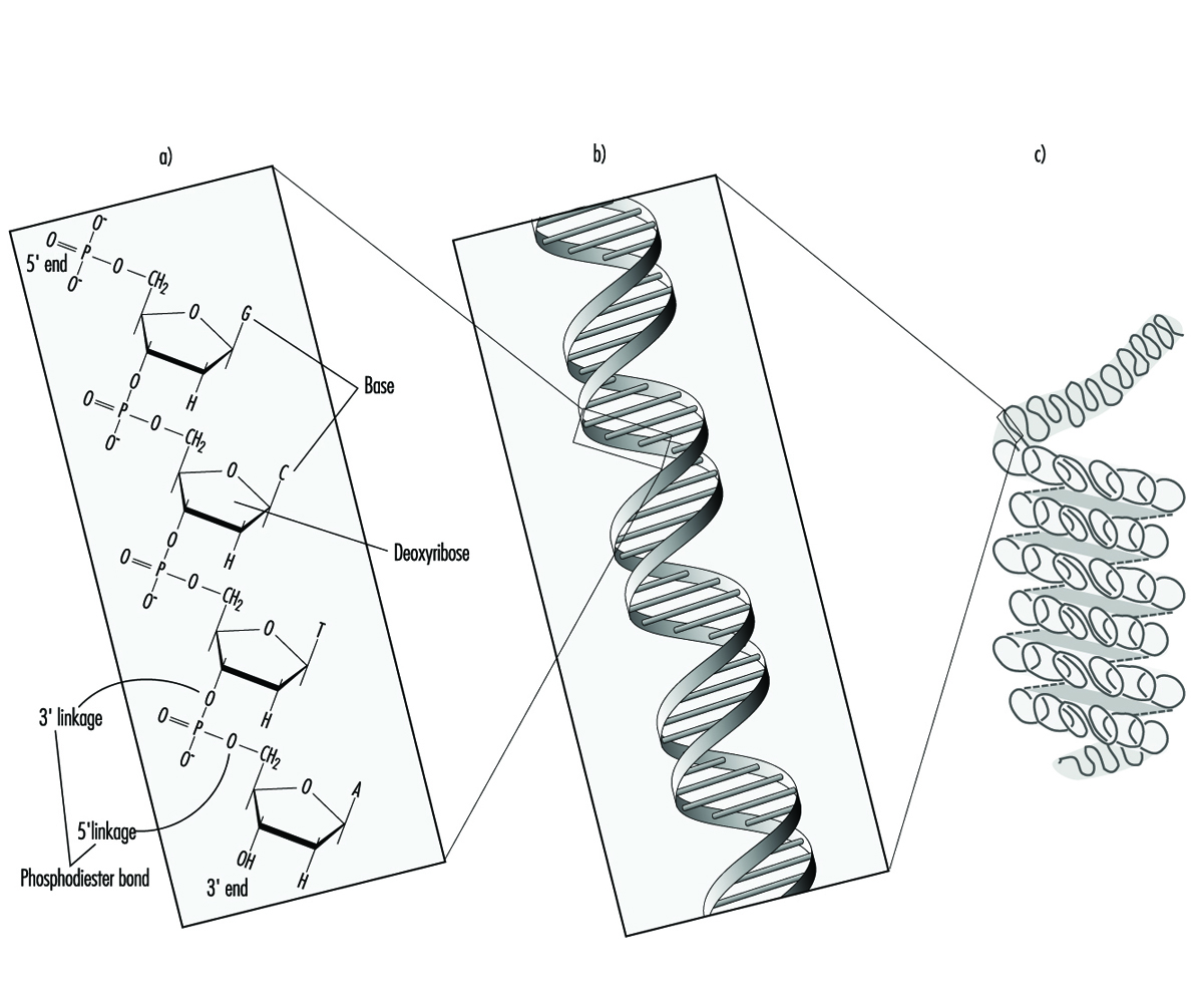
In higher organisms, hereditary information is organized in chromosomes. Chromosomes consist of tightly condensed strands of protein-associated DNA. Within a single chromosome, each DNA molecule exists as a pair of long, unbranched chains of nucleotide subunits linked together by phosphodiester bonds that join the 5 carbon of one deoxyribose moiety to the 3 carbon of the next (figure 1). In addition, one of four different nucleotide bases (adenine, cytosine, guanine or thymine) is attached to each deoxyribose subunit like beads on a string. Three-dimensionally, each pair of DNA strands forms a double helix with all of the bases oriented toward the inside of the spiral. Within the helix, each base is associated with its complementary base on the opposite DNA strand; hydrogen bonding dictates strong, noncovalent pairing of adenine with thymine and guanine with cytosine (figure 1). Since the sequence of nucleotide bases is complementary throughout the entire length of the duplex DNA molecule, both strands carry essentially the same genetic information. In fact, during DNA replication each strand serves as a template for the production of a new partner strand.

Figure 1. The (a) primary, (b) secondary and (c) tertiary organization of human hereditary information

TOX090F1Using RNA and an array of different proteins, the cell ultimately deciphers the information encoded by the linear sequence of bases within specific regions of DNA (genes) and produces proteins that are essential for basic cell survival as well as normal growth and differentiation. In essence, the nucleotides function like a biological alphabet which is used to code for amino acids, the building blocks of proteins.

When incorrect nucleotides are inserted or nucleotides are lost, or when unnecessary nucleotides are added during DNA synthesis, the mistake is called a mutation. It has been estimated that less than one mutation occurs for every 109 nucleotides incorporated during the normal replication of cells. Although mutations are not necessarily harmful, alterations causing inactivation or overexpression of important genes can result in a variety of disorders, including cancer, hereditary disease, developmental abnormalities, infertility and embryonic or perinatal death. Very rarely, a mutation can lead to enhanced survival; such occurrences are the basis of natural selection.

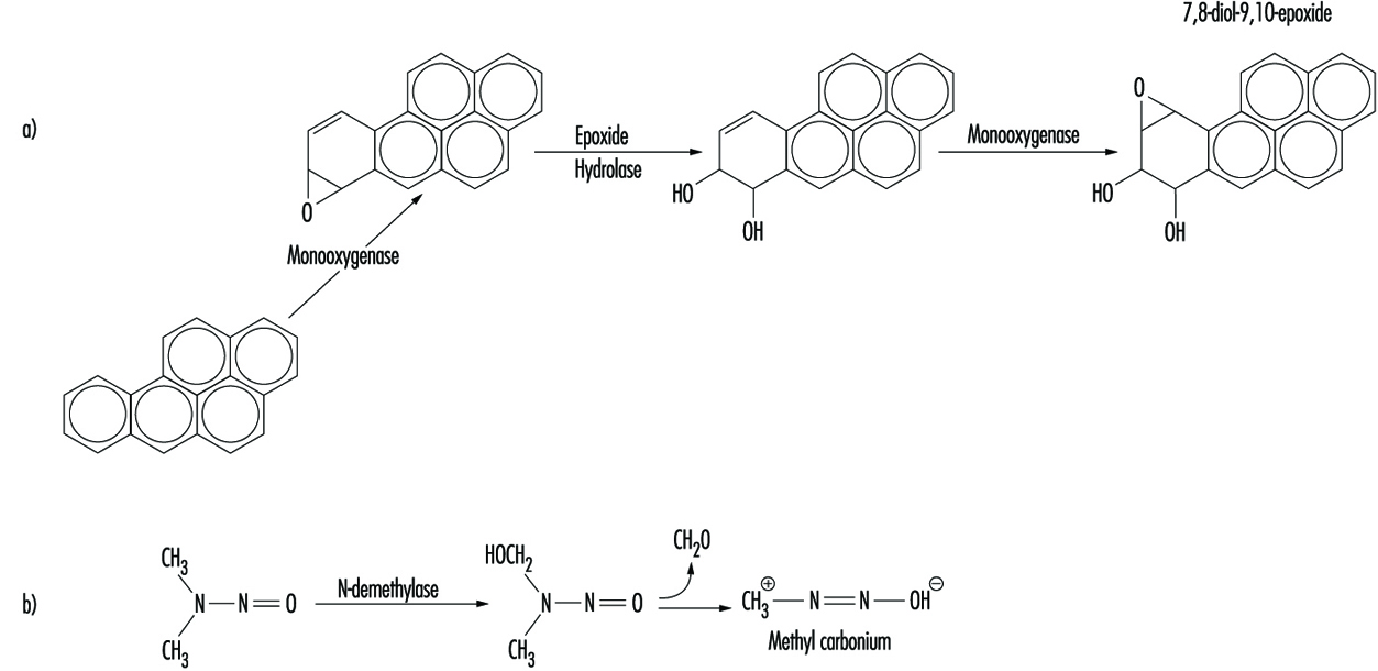
Although some chemicals react directly with DNA, most require metabolic activation. In the latter case, electrophilic intermediates such as epoxides or carbonium ions are ultimately responsible for inducing lesions at a variety of nucleophilic sites within the genetic material (figure 2). In other instances, genotoxicity is mediated by by-products of compound interaction with intracellular lipids, proteins, or oxygen.

Figure 2. Bioactivation of: a) benzo(a)pyrene; and b) N-nitrosodimethylamine

TOX090F2

Because of their relative abundance in cells, proteins are the most frequent target of toxicant interaction. However, modification of DNA is of greater concern due to the central role of this molecule in regulating growth and differentiation through multiple generations of cells.

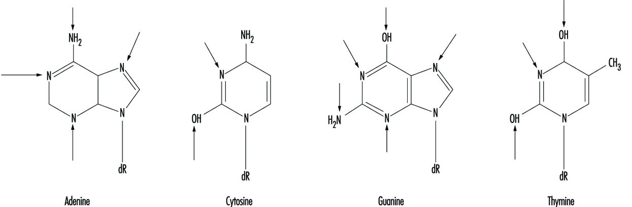
At the molecular level, electrophilic compounds tend to attack oxygen and nitrogen in DNA. The sites that are most prone to modification are illustrated in figure 3. Although oxygens within phosphate groups in the DNA backbone are also targets for chemical modification, damage to bases is thought to be biologically more relevant since these groups are considered to be the primary informational elements in the DNA molecule.

Figure 3. Primary sites of chemically-induced DNA damage

TOX090F3

Compounds that contain one electrophilic moiety typically exert genotoxicity by producing mono-adducts in DNA. Similarly, compounds that contain two or more reactive moieties can react with two different nucleophilic centres and thereby produce intra- or inter-molecular crosslinks in genetic material (figure 4). Interstrand DNA-DNA and DNA-protein crosslinks can be particularly cytotoxic since they can form complete blocks to DNA replication. For obvious reasons, the death of a cell eliminates the possibility that it will be mutated or neoplastically transformed. Genotoxic agents can also act by inducing breaks in the phosphodiester backbone, or between bases and sugars (producing abasic sites) in DNA. Such breaks may be a direct result of chemical reactivity at the damage site, or may occur during the repair of one of the aforementioned types of DNA lesion.

Figure 4. Various types of damage to the protein-DNA complex

TOX090F4

Over the past thirty to forty years, a variety of techniques have been developed to monitor the type of genetic damage induced by various chemicals. Such assays are described in detail elsewhere in this chapter and Encyclopaedia.

Misreplication of “microlesions” such as mono-adducts, abasic sites or single-strand breaks may ultimately result in nucleotide base-pair substitutions, or the insertion or deletion of short polynucleotide fragments in chromosomal DNA. In contrast, “macrolesions,” such as bulky adducts, crosslinks, or double-strand breaks may trigger the gain, loss or rearrangement of relatively large pieces of chromosomes. In any case, the consequences can be devastating to the organism since any one of these events can lead to cell death, loss of function or malignant transformation of cells. Exactly how DNA damage causes cancer is largely unknown. It is currently believed the process may involve inappropriate activation of proto-oncogenes such as myc and ras, and/or inactivation of recently identified tumour suppressor genes such as p53. Abnormal expression of either type of gene abrogates normal cellular mechanisms for controlling cell proliferation and/or differentiation.

The preponderance of experimental evidence indicates that the development of cancer following exposure to electrophilic compounds is a relatively rare event. This can be explained, in part, by the cell’s intrinsic ability to recognize and repair damaged DNA or the failure of cells with damaged DNA to survive. During repair, the damaged base, nucleotide or short stretch of nucleotides surrounding the damage site is removed and (using the opposite strand as a template) a new piece of DNA is synthesized and spliced into place. To be effective, DNA repair must occur with great accuracy prior to cell division, before opportunities for the propagation of mutation.

Clinical studies have shown that people with inherited defects in the ability to repair damaged DNA frequently develop cancer and/or developmental abnormalities at an early age (table 1). Such examples provide strong evidence linking accumulation of DNA damage to human disease. Similarly, agents that promote cell proliferation (such as tetradecanoylphorbol acetate) often enhance carcinogenesis. For these compounds, the increased likelihood of neoplastic transformation may be a direct consequence of a decrease in the time available for the cell to carry out adequate DNA repair.

Table 1. Hereditary, cancer-prone disorders that appear to involve defects in DNA repair

Syndrome Symptoms Cellular phenotype
Ataxia telangiectasia Neurological deterioration
Immunodeficiency
High incidence of lymphoma
Hypersensitivity to ionizing radiation and certain alkylating agents.
Dysregulated replication of damaged DNA (may indicate shortened time for DNA repair)
Bloom’s syndrome Developmental abnormalities
Lesions on exposed skin
High incidence of tumours of the immune system and gastrointestinal tract
High frequency of chromosomal aberrations
Defective ligation of breaks associated with DNA repair
Fanconi’s anaemia Growth retardation
High incidence of leukaemia
Hypersensitivity to crosslinking agents
High frequency of chromosomal aberrations
Defective repair of crosslinks in DNA
Hereditary nonpolyposis colon cancer High incidence of colon cancer Defect in DNA mismatch repair (when insertion of wrong nucleotide occurs during replication)
Xeroderma pigmentosum High incidence of epithelioma on exposed areas of skin
Neurological impairment (in many cases)
Hypersensitivity to UV light and many chemical carcinogens
Defects in excision repair and/or replication of damaged DNA


The earliest theories on how chemicals interact with DNA can be traced back to studies conducted during the development of mustard gas for use in warfare. Further understanding grew out of efforts to identify anticancer agents that would selectively arrest the replication of rapidly dividing tumour cells. Increased public concern over hazards in our environment has prompted additional research into the mechanisms and consequences of chemical interaction with the genetic material. Examples of various types of chemicals which exert genotoxicity are presented in table 2.

Table 2. Examples of chemicals that exhibit genotoxicity in human cells

Class of chemical Example Source of exposure Probable genotoxic lesion
Aflatoxins Aflatoxin B1 Contaminated food Bulky DNA adducts
Aromatic amines 2-Acetylaminofluorene Environmental Bulky DNA adducts
Aziridine quinones Mitomycin C Cancer chemotherapy Mono-adducts, interstrand crosslinks and single-strand breaks in DNA.
Chlorinated hydrocarbons Vinyl chloride Environmental Mono-adducts in DNA
Metals and metal compounds Cisplatin Cancer chemotherapy Both intra- and inter-strand crosslinks in DNA
  Nickel compounds Environmental Mono-adducts and single-strand breaks in DNA
Nitrogen mustards Cyclophosphamide Cancer chemotherapy Mono-adducts and interstrand crosslinks in DNA
Nitrosamines N-Nitrosodimethylamine Contaminated food Mono-adducts in DNA
Polycyclic aromatic hydrocarbons Benzo(a)pyrene Environmental Bulky DNA adducts


Back

Read 13298 times Last modified on Friday, 17 June 2022 21:40

" DISCLAIMER: The ILO does not take responsibility for content presented on this web portal that is presented in any language other than English, which is the language used for the initial production and peer-review of original content. Certain statistics have not been updated since the production of the 4th edition of the Encyclopaedia (1998)."

Contents

Toxicology References

Andersen, KE and HI Maibach. 1985. Contact allergy predictive tests on guinea pigs. Chap. 14 in Current Problems in Dermatology. Basel: Karger.

Ashby, J and RW Tennant. 1991. Definitive relationships among chemical structure, carcinogenicity and mutagenicity for 301 chemicals tested by the US NTP. Mutat Res 257:229-306.

Barlow, S and F Sullivan. 1982. Reproductive Hazards of Industrial Chemicals. London: Academic Press.

Barrett, JC. 1993a. Mechanisms of action of known human carcinogens. In Mechanisms of Carcinogenesis in Risk Identification, edited by H Vainio, PN Magee, DB McGregor, and AJ McMichael. Lyon: International Agency for Research on Cancer (IARC).

—. 1993b. Mechanisms of multistep carcinogenesis and carcinogen risk assessment. Environ Health Persp 100:9-20.

Bernstein, ME. 1984. Agents affecting the male reproductive system: Effects of structure on activity. Drug Metab Rev 15:941-996.

Beutler, E. 1992. The molecular biology of G6PD variants and other red cell defects. Annu Rev Med 43:47-59.

Bloom, AD. 1981. Guidelines for Reproductive Studies in Exposed Human Populations. White Plains, New York: March of Dimes Foundation.

Borghoff, S, B Short and J Swenberg. 1990. Biochemical mechanisms and pathobiology of a-2-globulin nephropathy. Annu Rev Pharmacol Toxicol 30:349.

Burchell, B, DW Nebert, DR Nelson, KW Bock, T Iyanagi, PLM Jansen, D Lancet, GJ Mulder, JR Chowdhury, G Siest, TR Tephly, and PI Mackenzie. 1991. The UPD-glucuronosyltransferase gene superfamily: Suggested nomenclature based on evolutionary divergence. DNA Cell Biol 10:487-494.

Burleson, G, A Munson, and J Dean. 1995. Modern Methods in Immunotoxicology. New York: Wiley.

Capecchi, M. 1994. Targeted gene replacement. Sci Am 270:52-59.

Carney, EW. 1994. An integrated perspective on the developmental toxicity of ethylene glycol. Rep Toxicol 8:99-113.

Dean, JH, MI Luster, AE Munson, and I Kimber. 1994. Immunotoxicology and Immunopharmacology. New York: Raven Press.

Descotes, J. 1986. Immunotoxicology of Drugs and Chemicals. Amsterdam: Elsevier.

Devary, Y, C Rosette, JA DiDonato, and M Karin. 1993. NFkB activation by ultraviolet light not dependent on a nuclear signal. Science 261:1442-1445.

Dixon, RL. 1985. Reproductive Toxicology. New York: Raven Press.

Duffus, JH. 1993. Glossary for chemists of terms used in toxicology. Pure Appl Chem 65:2003-2122.

Elsenhans, B, K Schuemann, and W Forth. 1991. Toxic metals: Interactions with essential metals. In Nutrition, Toxicity and Cancer, edited by IR Rowland. Boca-Raton: CRC Press.

Environmental Protection Agency (EPA). 1992. Guidelines for exposure assessment. Federal Reg 57:22888-22938.

—. 1993. Principles of neurotoxicity risk assessment. Federal Reg 58:41556-41598.

—. 1994. Guidelines for Reproductive Toxicity Assessment. Washington, DC: US EPA: Office of Research and Development.

Fergusson, JE. 1990. The Heavy Elements. Chap. 15 in Chemistry, Environmental Impact and Health Effects. Oxford: Pergamon.

Gehring, PJ, PG Watanabe, and GE Blau. 1976. Pharmacokinetic studies in evaluation of the toxicological and environmental hazard of chemicals. New Concepts Saf Eval 1(Part 1, Chapter 8):195-270.

Goldstein, JA and SMF de Morais. 1994. Biochemistry and molecular biology of the human CYP2C subfamily. Pharmacogenetics 4:285-299.

Gonzalez, FJ. 1992. Human cytochromes P450: Problems and prospects. Trends Pharmacol Sci 13:346-352.

Gonzalez, FJ, CL Crespi, and HV Gelboin. 1991. cDNA-expressed human cytochrome P450: A new age in molecular toxicology and human risk assessment. Mutat Res 247:113-127.

Gonzalez, FJ and DW Nebert. 1990. Evolution of the P450 gene superfamily: Animal-plant “warfare,” molecular drive, and human genetic differences in drug oxidation. Trends Genet 6:182-186.

Grant, DM. 1993. Molecular genetics of the N-acetyltransferases. Pharmacogenetics 3:45-50.

Gray, LE, J Ostby, R Sigmon, J Ferrel, R Linder, R Cooper, J Goldman, and J Laskey. 1988. The development of a protocol to assess reproductive effects of toxicants in the rat. Rep Toxicol 2:281-287.

Guengerich, FP. 1989. Polymorphism of cytochrome P450 in humans. Trends Pharmacol Sci 10:107-109.

—. 1993. Cytochrome P450 enzymes. Am Sci 81:440-447.

Hansch, C and A Leo. 1979. Substituent Constants for Correlation Analysis in Chemistry and Biology. New York: Wiley.

Hansch, C and L Zhang. 1993. Quantitative structure-activity relationships of cytochrome P450. Drug Metab Rev 25:1-48.

Hayes AW. 1988. Principles and Methods of Toxicology. 2nd ed. New York: Raven Press.

Heindell, JJ and RE Chapin. 1993. Methods in Toxicology: Male and Female Reproductive Toxicology. Vol. 1 and 2. San Diego, Calif.: Academic Press.

International Agency for Research on Cancer (IARC). 1992. Solar and ultraviolet radiation. Lyon: IARC.

—. 1993. Occupational Exposures of Hairdressers and Barbers and Personal Use of Hair Colourants: Some Hair Dyes, Cosmetic Colourants, Industrial Dyestuffs and Aromatic Amines. Lyon: IARC.

—. 1994a. Preamble. Lyon: IARC.

—. 1994b. Some Industrial Chemicals. Lyon: IARC.

International Commission on Radiological Protection (ICRP). 1965. Principles of Environmental Monitoring Related to the Handling of Radioactive Materials. Report of Committee IV of The International Commission On Radiological Protection. Oxford: Pergamon.

International Program on Chemical Safety (IPCS). 1991. Principles and Methods for the Assessment of Nephrotoxicity Associated With Exposure to Chemicals, EHC 119. Geneva: WHO.

—. 1996. Principles and Methods for Assessing Direct Immunotoxicity Associated With Exposure to Chemicals, EHC 180. Geneva: WHO.

Johanson, G and PH Naslund. 1988. Spreadsheet programming - a new approach in physiologically based modeling of solvent toxicokinetics. Toxicol Letters 41:115-127.

Johnson, BL. 1978. Prevention of Neurotoxic Illness in Working Populations. New York: Wiley.

Jones, JC, JM Ward, U Mohr, and RD Hunt. 1990. Hemopoietic System, ILSI Monograph, Berlin: Springer Verlag.

Kalow, W. 1962. Pharmocogenetics: Heredity and the Response to Drugs. Philadelphia: WB Saunders.

—. 1992. Pharmocogenetics of Drug Metabolism. New York: Pergamon.

Kammüller, ME, N Bloksma, and W Seinen. 1989. Autoimmunity and Toxicology. Immune Dysregulation Induced By Drugs and Chemicals. Amsterdam: Elsevier Sciences.

Kawajiri, K, J Watanabe, and SI Hayashi. 1994. Genetic polymorphism of P450 and human cancer. In Cytochrome P450: Biochemistry, Biophysics and Molecular Biology, edited by MC Lechner. Paris: John Libbey Eurotext.

Kehrer, JP. 1993. Free radicals as mediators of tissue injury and disease. Crit Rev Toxicol 23:21-48.

Kellerman, G, CR Shaw, and M Luyten-Kellerman. 1973. Aryl hydrocarbon hydroxylase inducibility and bronochogenic carcinoma. New Engl J Med 289:934-937.

Khera, KS. 1991. Chemically induced alterations maternal homeostasis and histology of conceptus: Their etiologic significance in rat fetal anomalies. Teratology 44:259-297.

Kimmel, CA, GL Kimmel, and V Frankos. 1986. Interagency Regulatory Liaison Group workshop on reproductive toxicity risk assessment. Environ Health Persp 66:193-221.

Klaassen, CD, MO Amdur and J Doull (eds.). 1991. Casarett and Doull´s Toxicology. New York: Pergamon Press.

Kramer, HJ, EJHM Jansen, MJ Zeilmaker, HJ van Kranen and ED Kroese. 1995. Quantitative methods in toxicology for human dose-response assessment. RIVM-report nr. 659101004.

Kress, S, C Sutter, PT Strickland, H Mukhtar, J Schweizer, and M Schwarz. 1992. Carcinogen-specific mutational pattern in the p53 gene in ultraviolet B radiation-induced squamous cell carcinomas of mouse skin. Cancer Res 52:6400-6403.

Krewski, D, D Gaylor, M Szyazkowicz. 1991. A model-free approach to low-dose extrapolation. Env H Pers 90:270-285.

Lawton, MP, T Cresteil, AA Elfarra, E Hodgson, J Ozols, RM Philpot, AE Rettie, DE Williams, JR Cashman, CT Dolphin, RN Hines, T Kimura, IR Phillips, LL Poulsen, EA Shephare, and DM Ziegler. 1994. A nomenclature for the mammalian flavin-containing monooxygenase gene family based on amino acid sequence identities. Arch Biochem Biophys 308:254-257.

Lewalter, J and U Korallus. 1985. Blood protein conjugates and acetylation of aromatic amines. New findings on biological monitoring. Int Arch Occup Environ Health 56:179-196.

Majno, G and I Joris. 1995. Apoptosis, oncosis, and necrosis: An overview of cell death. Am J Pathol 146:3-15.

Mattison, DR and PJ Thomford. 1989. The mechanism of action of reproductive toxicants. Toxicol Pathol 17:364-376.

Meyer, UA. 1994. Polymorphisms of cytochrome P450 CYP2D6 as a risk factor in carcinogenesis. In Cytochrome P450: Biochemistry, Biophysics and Molecular Biology, edited by MC Lechner. Paris: John Libbey Eurotext.

Moller, H, H Vainio and E Heseltine. 1994. Quantitative estimation and prediction of risk at the International Agency for Research on Cancer. Cancer Res 54:3625-3627.

Moolenaar, RJ. 1994. Default assumptions in carcinogen risk assessment used by regulatory agencies. Regul Toxicol Pharmacol 20:135-141.

Moser, VC. 1990. Screening approaches to neurotoxicity: A functional observational battery. J Am Coll Toxicol 1:85-93.

National Research Council (NRC). 1983. Risk Assessment in the Federal Government: Managing the Process. Washington, DC: NAS Press.

—. 1989. Biological Markers in Reproductive Toxicity. Washington, DC: NAS Press.

—. 1992. Biologic Markers in Immunotoxicology. Subcommittee on Toxicology. Washington, DC: NAS Press.

Nebert, DW. 1988. Genes encoding drug-metabolizing enzymes: Possible role in human disease. In Phenotypic Variation in Populations, edited by AD Woodhead, MA Bender, and RC Leonard. New York: Plenum Publishing.

—. 1994. Drug-metabolizing enzymes in ligand-modulated transcription. Biochem Pharmacol 47:25-37.

Nebert, DW and WW Weber. 1990. Pharmacogenetics. In Principles of Drug Action. The Basis of Pharmacology, edited by WB Pratt and PW Taylor. New York: Churchill-Livingstone.

Nebert, DW and DR Nelson. 1991. P450 gene nomenclature based on evolution. In Methods of Enzymology. Cytochrome P450, edited by MR Waterman and EF Johnson. Orlando, Fla: Academic Press.

Nebert, DW and RA McKinnon. 1994. Cytochrome P450: Evolution and functional diversity. Prog Liv Dis 12:63-97.

Nebert, DW, M Adesnik, MJ Coon, RW Estabrook, FJ Gonzalez, FP Guengerich, IC Gunsalus, EF Johnson, B Kemper, W Levin, IR Phillips, R Sato, and MR Waterman. 1987. The P450 gene superfamily: Recommended nomenclature. DNA Cell Biol 6:1-11.

Nebert, DW, DR Nelson, MJ Coon, RW Estabrook, R Feyereisen, Y Fujii-Kuriyama, FJ Gonzalez, FP Guengerich, IC Gunsalas, EF Johnson, JC Loper, R Sato, MR Waterman, and DJ Waxman. 1991. The P450 superfamily: Update on new sequences, gene mapping, and recommended nomenclature. DNA Cell Biol 10:1-14.

Nebert, DW, DD Petersen, and A Puga. 1991. Human AH locus polymorphism and cancer: Inducibility of CYP1A1 and other genes by combustion products and dioxin. Pharmacogenetics 1:68-78.

Nebert, DW, A Puga, and V Vasiliou. 1993. Role of the Ah receptor and the dioxin-inducible [Ah] gene battery in toxicity, cancer, and signal transduction. Ann NY Acad Sci 685:624-640.

Nelson, DR, T Kamataki, DJ Waxman, FP Guengerich, RW Estabrook, R Feyereisen, FJ Gonzalez, MJ Coon, IC Gunsalus, O Gotoh, DW Nebert, and K Okuda. 1993. The P450 superfamily: Update on new sequences, gene mapping, accession numbers, early trivial names of enzymes, and nomenclature. DNA Cell Biol 12:1-51.

Nicholson, DW, A All, NA Thornberry, JP Vaillancourt, CK Ding, M Gallant, Y Gareau, PR Griffin, M Labelle, YA Lazebnik, NA Munday, SM Raju, ME Smulson, TT Yamin, VL Yu, and DK Miller. 1995. Identification and inhibition of the ICE/CED-3 protease necessary for mammalian apoptosis. Nature 376:37-43.

Nolan, RJ, WT Stott, and PG Watanabe. 1995. Toxicologic data in chemical safety evaluation. Chap. 2 in Patty’s Industrial Hygiene and Toxicology, edited by LJ Cralley, LV Cralley, and JS Bus. New York: John Wiley & Sons.

Nordberg, GF. 1976. Effect and Dose-Response Relationships of Toxic Metals. Amsterdam: Elsevier.

Office of Technology Assessment (OTA). 1985. Reproductive Hazards in the Workplace. Document No. OTA-BA-266. Washington, DC: Government Printing Office.

—. 1990. Neurotoxicity: Identifying and Controlling Poisons of the Nervous System. Document No. OTA-BA-436. Washington, DC: Government Printing Office.

Organization for Economic Cooperation and Development (OECD). 1993. US EPA/EC Joint Project On the Evaluation of (Quantitative) Structure Activity Relationships. Paris: OECD.

Park, CN and NC Hawkins. 1993. Technology review; an overview of cancer risk assessment. Toxicol Methods 3:63-86.

Pease, W, J Vandenberg, and WK Hooper. 1991. Comparing alternative approaches to establishing regulatory levels for reproductive toxicants: DBCP as a case study. Environ Health Persp 91:141-155.

Prpi<F"WP MultinationalA Roman"P6.5>ƒ<F255P255>-Maji<F"WP MultinationalA Roman"P6.5%0>ƒ<F255P255>, D, S Telišman, and S Kezi<F"WP MultinationalA Roman"P6.5%0>ƒ<F255P255>. 1984. In vitro study on lead and alcohol interaction and the inhibition of erythrocyte delta-aminolevulinic acid dehydratase in man. Scand J Work Environ Health 10:235-238.

Reitz, RH, RJ Nolan, and AM Schumann. 1987. Development of multispecies, multiroute pharmacokinetic models for methylene chloride and 1,1,1-trichloroethane. In Pharmacokinetics and Risk Assessment, Drinking Water and Health. Washington, DC: National Academy Press.

Roitt, I, J Brostoff, and D Male. 1989. Immunology. London: Gower Medical Publishing.

Sato, A. 1991. The effect of environmental factors on the pharmacokinetic behaviour of organic solvent vapours. Ann Occup Hyg 35:525-541.

Silbergeld, EK. 1990. Developing formal risk assessment methods for neurotoxicants: An evaluation of the state of the art. In Advances in Neurobehavioral Toxicology, edited by BL Johnson, WK Anger, A Durao, and C Xintaras. Chelsea, Mich.: Lewis.

Spencer, PS and HH Schaumberg. 1980. Experimental and Clinical Neurotoxicology. Baltimore: Williams & Wilkins.

Sweeney, AM, MR Meyer, JH Aarons, JL Mills, and RE LePorte. 1988. Evaluation of methods for the prospective identification of early fetal losses in environmental epidemiology studies. Am J Epidemiol 127:843-850.

Taylor, BA, HJ Heiniger, and H Meier. 1973. Genetic analysis of resistance to cadmium-induced testicular damage in mice. Proc Soc Exp Biol Med 143:629-633.

Telišman, S. 1995. Interactions of essential and/or toxic metals and metalloids regarding interindividual differences in susceptibility to various toxicants and chronic diseases in man. Arh rig rada toksikol 46:459-476.

Telišman, S, A Pinent, and D Prpi<F"WP MultinationalA Roman"P6.5J255%0>ƒ<F255P255J0>-Maji<F"WP MultinationalA Roman"P6.5J255%0>ƒ<F255P255J0>. 1993. Lead interference in zinc metabolism and the lead and zinc interaction in humans as a possible explanation of apparent individual susceptibility to lead. In Heavy Metals in the Environment, edited by RJ Allan and JO Nriagu. Edinburgh: CEP Consultants.

Telišman, S, D Prpi<F"WP MultinationalA Roman"P6.5%0>ƒ<F255P255>-Maji<F"WP MultinationalA Roman"P6.5%0>ƒ<F255P255>, and S Kezi<F"WP MultinationalA Roman"P6.5%0>ƒ<F255P255>. 1984. In vivo study on lead and alcohol interaction and the inhibition of erythrocyte delta-aminolevulinic acid dehydratase in man. Scand J Work Environ Health 10:239-244.

Tilson, HA and PA Cabe. 1978. Strategies for the assessment of neurobehavioral consequences of environmental factors. Environ Health Persp 26:287-299.

Trump, BF and AU Arstila. 1971. Cell injury and cell death. In Principles of Pathobiology, edited by MF LaVia and RB Hill Jr. New York: Oxford Univ. Press.

Trump, BF and IK Berezesky. 1992. The role of cytosolic Ca2<F"Symbol"P8>+<F255P255> in cell injury, necrosis and apoptosis. Curr Opin Cell Biol 4:227-232.

—. 1995. Calcium-mediated cell injury and cell death. FASEB J 9:219-228.

Trump, BF, IK Berezesky, and A Osornio-Vargas. 1981. Cell death and the disease process. The role of cell calcium. In Cell Death in Biology and Pathology, edited by ID Bowen and RA Lockshin. London: Chapman & Hall.

Vos, JG, M Younes and E Smith. 1995. Allergic Hyper-sensitivities Induced by Chemicals: Recommendations for Prevention Published on Behalf of the World Health Organization Regional Office for Europe. Boca Raton, FL: CRC Press.

Weber, WW. 1987. The Acetylator Genes and Drug Response. New York: Oxford Univ. Press.

World Health Organization (WHO). 1980. Recommended Health-Based Limits in Occupational Exposure to Heavy Metals. Technical Report Series, No. 647. Geneva: WHO.

—. 1986. Principles and Methods for the Assessment of Neurotoxicity Associated With Exposure to Chemicals. Environmental Health Criteria, No.60. Geneva: WHO.

—. 1987. Air Quality Guidelines for Europe. European Series, No. 23. Copenhagen: WHO Regional Publications.

—. 1989. Glossary of Terms On Chemical Safety for Use in IPCS Publications. Geneva: WHO.

—. 1993. The Derivation of Guidance Values for Health-Based Exposure Limits. Environmental Health Criteria, unedited draft. Geneva: WHO.

Wyllie, AH, JFR Kerr, and AR Currie. 1980. Cell death: The significance of apoptosis. Int Rev Cytol 68:251-306.

@REFS LABEL = Other relevant readings

Albert, RE. 1994. Carcinogen risk assessment in the US Environmental Protection Agency. Crit. Rev. Toxicol 24:75-85.

Alberts, B, D Bray, J Lewis, M Raff, K Roberts, and JD Watson. 1988. Molecular Biology of the Cell. New York: Garland Publishing.

Ariens, EJ. 1964. Molecular Pharmacology. Vol.1. New York: Academic Press.

Ariens, EJ, E Mutschler, and AM Simonis. 1978. Allgemeine Toxicologie [General Toxicology]. Stuttgart: Georg Thieme Verlag.

Ashby, J and RW Tennant. 1994. Prediction of rodent carcinogenicity for 44 chemicals: Results. Mutagenesis 9:7-15.

Ashford, NA, CJ Spadafor, DB Hattis, and CC Caldart. 1990. Monitoring the Worker for Exposure and Disease. Baltimore: Johns Hopkins Univ. Press.

Balabuha, NS and GE Fradkin. 1958. Nakoplenie radioaktivnih elementov v organizme I ih vivedenie [Accumulation of Radioactive Elements in the Organism and their Excretion]. Moskva: Medgiz.

Balls, M, J Bridges, and J Southee. 1991. Animals and Alternatives in Toxicology Present Status and Future Prospects. Nottingham, UK: The Fund for Replacement of Animals in Medical Experiments.

Berlin, A, J Dean, MH Draper, EMB Smith, and F Spreafico. 1987. Immunotoxicology. Dordrecht: Martinus Nijhoff.

Boyhous, A. 1974. Breathing. New York: Grune & Stratton.

Brandau, R and BH Lippold. 1982. Dermal and Transdermal Absorption. Stuttgart: Wissenschaftliche Verlagsgesellschaft.

Brusick, DJ. 1994. Methods for Genetic Risk Assessment. Boca Raton: Lewis Publishers.

Burrell, R. 1993. Human immune toxicity. Mol Aspects Med 14:1-81.

Castell, JV and MJ Gómez-Lechón. 1992. In Vitro Alternatives to Animal Pharmaco-Toxicology. Madrid, Spain: Farmaindustria.

Chapman, G. 1967. Body Fluids and their Functions. London: Edward Arnold.

Committee on Biological Markers of the National Research Council. 1987. Biological markers in environmental health research. Environ Health Persp 74:3-9.

Cralley, LJ, LV Cralley and JS Bus (eds.). 1978. Patty’s Industrial Hygiene and Toxicology. New York: Witey.

Dayan, AD, RF Hertel, E Heseltine, G Kazantis, EM Smith, and MT Van der Venne. 1990. Immunotoxicity of Metals and Immunotoxicology. New York: Plenum Press.

Djuric, D. 1987. Molecular-cellular Aspects of Occupational Exposure to Toxic Chemicals. In Part 1 Toxicokinetics. Geneva: WHO.

Duffus, JH. 1980. Environmental Toxicology. London: Edward Arnold.

ECOTOC. 1986. Structure-Activity Relationship in Toxicology and Ecotoxicology, Monograph No. 8. Brussels: ECOTOC.

Forth, W, D Henschler, and W Rummel. 1983. Pharmakologie und Toxikologie. Mannheim: Biblio- graphische Institut.

Frazier, JM. 1990. Scientific criteria for Validation of in VitroToxicity Tests. OECD Environmental Monograph, no. 36. Paris: OECD.

—. 1992. In Vitro Toxicity—Applications to Safety Evaluation. New York: Marcel Dekker.

Gad, SC. 1994. In Vitro Toxicology. New York: Raven Press.

Gadaskina, ID. 1970. Zhiroraya tkan I yadi [Fatty Tissues and Toxicants]. In Aktualnie Vaprosi promishlenoi toksikolgii [Actual Problems in Occupational Toxicology], edited by NV Lazarev. Leningrad: Ministry of Health RSFSR.

Gaylor, DW. 1983. The use of safety factors for controlling risk. J Toxicol Environ Health 11:329-336.

Gibson, GG, R Hubbard, and DV Parke. 1983. Immunotoxicology. London: Academic Press.

Goldberg, AM. 1983-1995. Alternatives in Toxicology. Vol. 1-12. New York: Mary Ann Liebert.

Grandjean, P. 1992. Individual susceptibility to toxicity. Toxicol Letters 64/65:43-51.

Hanke, J and JK Piotrowski. 1984. Biochemyczne podstawy toksikologii [Biochemical Basis of Toxicology]. Warsaw: PZWL.

Hatch, T and P Gross. 1954. Pulmonary Deposition and Retention of Inhaled Aerosols. New York: Academic Press.

Health Council of the Netherlands: Committee on the Evaluation of the Carcinogenicity of Chemical Substances. 1994. Risk assessment of carcinogenic chemicals in The Netherlands. Regul Toxicol Pharmacol 19:14-30.

Holland, WC, RL Klein, and AH Briggs. 1967. Molekulaere Pharmakologie.

Huff, JE. 1993. Chemicals and cancer in humans: First evidence in experimental animals. Environ Health Persp 100:201-210.

Klaassen, CD and DL Eaton. 1991. Principles of toxicology. Chap. 2 in Casarett and Doull’s Toxicology, edited by CD Klaassen, MO Amdur and J Doull. New York: Pergamon Press.

Kossover, EM. 1962. Molecular Biochemistry. New York: McGraw-Hill.

Kundiev, YI. 1975.Vssavanie pesticidov cherez kozsu I profilaktika otravlenii [Absorption of Pesticides Through Skin and Prevention of Intoxication]. Kiev: Zdorovia.

Kustov, VV, LA Tiunov, and JA Vasiljev. 1975. Komvinovanie deistvie promishlenih yadov [Combined Effects of Industrial Toxicants]. Moskva: Medicina.

Lauwerys, R. 1982. Toxicologie industrielle et intoxications professionelles. Paris: Masson.

Li, AP and RH Heflich. 1991. Genetic Toxicology. Boca Raton: CRC Press.

Loewey, AG and P Siekewitz. 1969. Cell Structure and Functions. New York: Holt, Reinhart and Winston.

Loomis, TA. 1976. Essentials of Toxicology. Philadelphia: Lea & Febiger.

Mendelsohn, ML and RJ Albertini. 1990. Mutation and the Environment, Parts A-E. New York: Wiley Liss.

Mettzler, DE. 1977. Biochemistry. New York: Academic Press.

Miller, K, JL Turk, and S Nicklin. 1992. Principles and Practice of Immunotoxicology. Oxford: Blackwells Scientific.

Ministry of International Trade and Industry. 1981. Handbook of Existing Chemical Substances. Tokyo: Chemical Daily Press.

—. 1987. Application for Approval of Chemicals by Chemical Substances Control Law. (In Japanese and in English). Tokyo: Kagaku Kogyo Nippo Press.

Montagna, W. 1956. The Structure and Function of Skin. New York: Academic Press.

Moolenaar, RJ. 1994. Carcinogen risk assessment: international comparison. Regul Toxicol Pharmacol 20:302-336.

National Research Council. 1989. Biological Markers in Reproductive Toxicity. Washington, DC: NAS Press.

Neuman, WG and M Neuman. 1958. The Chemical Dynamic of Bone Minerals. Chicago: The Univ. of Chicago Press.

Newcombe, DS, NR Rose, and JC Bloom. 1992. Clinical Immunotoxicology. New York: Raven Press.

Pacheco, H. 1973. La pharmacologie moleculaire. Paris: Presse Universitaire.

Piotrowski, JK. 1971. The Application of Metabolic and Excretory Kinetics to Problems of Industrial Toxicology. Washington, DC: US Department of Health, Education and Welfare.

—. 1983. Biochemical interactions of heavy metals: Methalothionein. In Health Effects of Combined Exposure to Chemicals. Copenhagen: WHO Regional Office for Europe.

Proceedings of the Arnold O. Beckman/IFCC Conference of Environmental Toxicology Biomarkers of Chemical Exposure. 1994. Clin Chem 40(7B).

Russell, WMS and RL Burch. 1959. The Principles of Humane Experimental Technique. London: Methuen & Co. Reprinted by Universities Federation for Animal Welfare,1993.

Rycroft, RJG, T Menné, PJ Frosch, and C Benezra. 1992. Textbook of Contact Dermatitis. Berlin: Springer-Verlag.

Schubert, J. 1951. Estimating radioelements in exposed individuals. Nucleonics 8:13-28.

Shelby, MD and E Zeiger. 1990. Activity of human carcinogens in the Salmonella and rodent bone-marrow cytogenetics tests. Mutat Res 234:257-261.

Stone, R. 1995. A molecular approach to cancer risk. Science 268:356-357.

Teisinger, J. 1984. Expositiontest in der Industrietoxikologie [Exposure Tests in Industrial Toxicology]. Berlin: VEB Verlag Volk und Gesundheit.

US Congress. 1990. Genetic Monitoring and Screening in the Workplace, OTA-BA-455. Washington, DC: US Government Printing Office.

VEB. 1981. Kleine Enzyklopaedie: Leben [Life]. Leipzig: VEB Bibliographische Institut.

Weil, E. 1975. Elements de toxicologie industrielle [Elements of Industrial Toxicology]. Paris: Masson et Cie.

World Health Organization (WHO). 1975. Methods Used in USSR for Establishing Safe Levels of Toxic Substances. Geneva: WHO.

1978. Principles and Methods for Evaluating the Toxicity of Chemicals, Part 1. Environmental Health Criteria, no.6. Geneva: WHO.

—. 1981. Combined Exposure to Chemicals, Interim Document no.11. Copenhagen: WHO Regional Office for Europe.

—. 1986. Principles of Toxicokinetic Studies. Environmental Health Criteria, no. 57. Geneva: WHO.

Yoftrey, JM and FC Courtice. 1956. Limphatics, Lymph and Lymphoid Tissue. Cambridge: Harvard Univ. Press.

Zakutinskiy, DI. 1959. Voprosi toksikologii radioaktivnih veshchestv [Problems of Toxicology of Radioactive Materials]. Moscow: Medgiz.

Zurlo, J, D Rudacille, and AM Goldberg. 1993. Animals and Alternatives in Testing: History, Science and Ethics. New York: Mary Ann Liebert.