Tuesday, 15 March 2011 14:46

The Electromagnetic Spectrum: Basic Physical Characteristics

Rate this item
(4 votes)

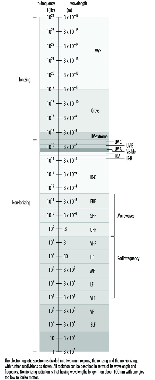
The most familiar form of electromagnetic energy is sunlight. The frequency of sunlight (visible light) is the dividing line between the more potent, ionizing radiation (x rays, cosmic rays) at higher frequencies and the more benign, non-ionizing radiation at lower frequencies. There is a spectrum of non-ionizing radiation. Within the context of this chapter, at the high end just below visible light is infrared radiation. Below that is the broad range of radio frequencies, which includes (in descending order) microwaves, cellular radio, television, FM radio and AM radio, short waves used in dielectric and induction heaters and, at the low end, fields with power frequency. The electromagnetic spectrum is illustrated in figure 1. 

Figure 1. The electromagnetic spectrum

ELF010F1

Just as visible light or sound permeates our environment, the space where we live and work, so do the energies of electromagnetic fields. Also, just as most of the sound energy we are exposed to is created by human activity, so too are the electromagnetic energies: from the weak levels emitted from our everyday electrical appliances—those that make our radio and TV sets work—to the high levels that medical practitioners apply for beneficial purposes—for example, diathermy (heat treatments). In general, the strength of such energies decreases rapidly with distance from the source. Natural levels of these fields in the environment are low.

Non-ionizing radiation (NIR) incorporates all radiation and fields of the electromagnetic spectrum that do not have enough energy to produce ionization of matter. That is, NIR is incapable of imparting enough energy to a molecule or atom to disrupt its structure by removing one or more electrons. The borderline between NIR and ionizing radiation is usually set at a wavelength of approximately 100 nanometres.

As with any form of energy, NIR energy has the potential to interact with biological systems, and the outcome may be of no significance, may be harmful in different degrees, or may be beneficial. With radiofrequency (RF) and microwave radiation, the main interaction mechanism is heating, but in the low-frequency part of the spectrum, fields of high intensity may induce currents in the body and thereby be hazardous. The interaction mechanisms for low-level field strengths are, however, unknown.


Quantities and Units

Fields at frequencies below about 300 MHz are quantified in terms of electric field strength (E) and magnetic field strength (H). E is expressed in volts per metre (V/m) and H in amperes per metre (A/m). Both are vector fields—that is, they are characterized by magnitude and direction at each point. For the low-frequency range the magnetic field is often expressed in terms of the flux density, B, with the SI unit tesla (T). When the fields in our daily environment are discussed, the subunit microtesla (μT) is usually the preferred unit. In some literature the flux density is expressed in gauss (G), and the conversion between these units is (for fields in air):

1 T = 104 G or 0.1 μT = 1 mG and 1 A/m = 1.26 μT.

Reviews of concepts, quantities, units and terminology for non-ionizing radiation protection, including radiofrequency radiation, are available (NCRP 1981; Polk and Postow 1986; WHO 1993).

The term radiation simply means energy transmitted by waves. Electromagnetic waves are waves of electric and magnetic forces, where a wave motion is defined as propagation of disturbances in a physical system. A change in the electric field is accompanied by a change in the magnetic field, and vice versa. These phenomena were described in 1865 by J.C. Maxwell in four equations which have come to be known as Maxwell’s Equations.

Electromagnetic waves are characterized by a set of parameters that include frequency (f), wavelength (λ), electric field strength, magnetic field strength, electric polarization (P) (the direction of the E field), velocity of propagation (c) and Poynting vector (S). Figure 2  illustrates the propagation of an electromagnetic wave in free space. The frequency is defined as the number of complete changes of the electric or magnetic field at a given point per second, and is expressed in hertz (Hz). The wavelength is the distance between two consecutive crests or troughs of the wave (maxima or minima). The frequency, wavelength and wave velocity (v) are interrelated as follows:

v = f λ

Figure 2. A plane wave propagating with the speed of light in the x-direction

ELF010F2

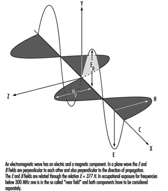
The velocity of an electromagnetic wave in free space is equal to the velocity of light, but the velocity in materials depends on the electrical properties of the material—that is, on its permittivity (ε) and permeability (μ). The permittivity concerns the material interactions with the electric field, and the permeability expresses the interactions with the magnetic field. Biological substances have permittivities that differ vastly from that of free space, being dependant on wavelength (especially in the RF range) and tissue type. The permeability of biological substances, however, is equal to that of free space.

In a plane wave, as illustrated in figure 2 , the electric field is perpendicular to the magnetic field and the direction of propagation is perpendicular to both the electric and the magnetic fields.


For a plane wave, the ratio of the value of the electric field strength to the value of the magnetic field strength, which is constant, is known as the characteristic impedance (Z):

Z = E/H

In free space, Z= 120π ≈ 377Ω but otherwise Z depends on the permittivity and permeability of the material the wave is travelling through.

Energy transfer is described by the Poynting vector, which represents the magnitude and direction of the electromagnetic flux density:

S = E x H

For a propagating wave, the integral of S over any surface represents the instantaneous power transmitted through this surface (power density). The magnitude of the Poynting vector is expressed in watts per square metre (W/m2) (in some literature the unit mW/cm2 is used—the conversion to SI units is 1 mW/cm2 = 10 W/m2) and for plane waves is related to the values of the electric and magnetic field strengths:

S = E2 / 120π = E2 / 377

and

S =120π H2 = 377 H2

Not all exposure conditions encountered in practice can be represented by plane waves. At distances close to sources of radio-frequency radiation the relationships characteristic of plane waves are not satisfied. The electromagnetic field radiated by an antenna can be divided into two regions: the near-field zone and the far-field zone. The boundary between these zones is usually put at:

r = 2a2 / λ

where a is the greatest dimension of the antenna.

In the near-field zone, exposure has to be characterized by both the electric and the magnetic fields. In the far-field one of these suffices, as they are interrelated by the above equations involving E and H. In practice, the near-field situation is often realized at frequencies below 300 Mhz.

Exposure to RF fields is further complicated by interactions of electromagnetic waves with objects. In general, when electromagnetic waves encounter an object some of the incident energy is reflected, some is absorbed and some is transmitted. The proportions of energy transmitted, absorbed or reflected by the object depend on the frequency and polarization of the field and the electrical properties and shape of the object. A superimposition of the incident and reflected waves results in standing waves and spatially non-uniform field distribution. Since waves are totally reflected from metallic objects, standing waves form close to such objects.

Since the interaction of RF fields with biological systems depends on many different field characteristics and the fields encountered in practice are complex, the following factors should be considered in describing exposures to RF fields:

  • whether exposure occurs in the near- or far-field zone
  • if near-field, then values for both E and H are needed; if far-field, then either E or H
  • spatial variation of the magnitude of the field(s)
  • field polarization, that is, the direction of the electric field with respect to the direction of wave propagation.


For exposure to low-frequency magnetic fields it is still not clear whether the field strength or flux density is the only important consideration. It may turn out that other factors are also important, such as the exposure time or the rapidity of the field changes.

The term electromagnetic field (EMF), as it is used in the news media and popular press, usually refers to electric and magnetic fields at the low-frequency end of the spectrum, but it can also be used in a much broader sense to include the whole spectrum of electromagnetic radiation. Note that in the low-frequency range the E and B fields are not coupled or interrelated in the same way that they are at higher frequencies, and it is therefore more accurate to refer to them as “electric and magnetic fields” rather than EMFs.


Back

Read 13134 times Last modified on Wednesday, 17 August 2011 17:44

" DISCLAIMER: The ILO does not take responsibility for content presented on this web portal that is presented in any language other than English, which is the language used for the initial production and peer-review of original content. Certain statistics have not been updated since the production of the 4th edition of the Encyclopaedia (1998)."

Contents

Radiation: Non-Ionizng References

Allen, SG. 1991. Radiofrequency field measurements and hazard assessment. J Radiol Protect 11:49-62.

American Conference of Governmental Industrial Hygienists (ACGIH). 1992. Documentation for the Threshold Limit Values. Cincinnati, Ohio: ACGIH.

—. 1993. Threshold Limit Values for Chemical Substances and Physical Agents and Biological Exposure Indices. Cincinnati, Ohio: ACGIH.

—. 1994a. Annual Report of ACGIH Physical Agents Threshold Limit Values Committee. Cincinnati, Ohio: ACGIH.

—. 1994b. TLV’s, Threshold Limit Values and Biological Exposure Indices for 1994-1995. Cincinnati, Ohio: ACGIH.

—. 1995. 1995-1996 Threshhold Limit Values for Chemical Substances and Physical Agents and Biological Exposure Indices. Cincinnati, Ohio: ACGIH.

—. 1996. TLVs© and BEIs©. Threshold Limit Values for Chemical Substances and Physical Agents; Biological Exposure Indices. Cincinnati, Ohio: ACGIH.

American National Standards Institute (ANSI). 1993. Safe Use of Lasers. Standard No. Z-136.1. New York: ANSI.

Aniolczyk, R. 1981. Measurements of hygienic evaluation of electromagnetic fields in the environment of diathermy, welders, and induction heaters. Medycina Pracy 32:119-128.

Bassett, CAL, SN Mitchell, and SR Gaston. 1982. Pulsing electromagnetic field treatment in ununited fractures and failed artrodeses. J Am Med Assoc 247:623-628.

Bassett, CAL, RJ Pawluk, and AA Pilla. 1974. Augmentation of bone repair by inductively coupled electromagnetic fields. Science 184:575-577.

Berger, D, F Urbach, and RE Davies. 1968. The action spectrum of erythema induced by ultraviolet radiation. In Preliminary Report XIII. Congressus Internationalis Dermatologiae, Munchen, edited by W Jadassohn and CG Schirren. New York: Springer-Verlag.

Bernhardt, JH. 1988a. The establishment of frequency dependent limits for electric and magnetic fields and evaluation of indirect effects. Rad Envir Biophys 27:1.

Bernhardt, JH and R Matthes. 1992. ELF and RF electromagnetic sources. In Non-Ionizing Radiation Protection, edited by MW Greene. Vancouver: UBC Press.

Bini, M, A Checcucci, A Ignesti, L Millanta, R Olmi, N Rubino, and R Vanni. 1986. Exposure of workers to intense RF electric fields that leak from plastic sealers. J Microwave Power 21:33-40.

Buhr, E, E Sutter, and Dutch Health Council. 1989. Dynamic filters for protective devices. In Dosimetry of Laser Radiation in Medicine and Biology, edited by GJ Mueller and DH Sliney. Bellingham, Wash: SPIE.

Bureau of Radiological Health. 1981. An Evaluation of Radiation Emission from Video Display Terminals. Rockville, MD: Bureau of Radiological Health.

Cleuet, A and A Mayer. 1980. Risques liés à l’utilisation industrielle des lasers. In Institut National de Recherche et de Sécurité, Cahiers de Notes Documentaires, No. 99 Paris: Institut National de Recherche et de Sécurité.

Coblentz, WR, R Stair, and JM Hogue. 1931. The spectral erythemic relation of the skin to ultraviolet radiation. In Proceedings of the National Academy of Sciences of the United States of America Washington, DC: National Academy of Sciences.

Cole, CA, DF Forbes, and PD Davies. 1986. An action spectrum for UV photocarcinogenesis. Photochem Photobiol 43(3):275-284.

Commission Internationale de L’Eclairage (CIE). 1987. International Lighting Vocabulary. Vienna: CIE.

Cullen, AP, BR Chou, MG Hall, and SE Jany. 1984. Ultraviolet-B damages corneal endothelium. Am J Optom Phys Opt 61(7):473-478.

Duchene, A, J Lakey, and M Repacholi. 1991. IRPA Guidelines On Protection Against Non-Ionizing Radiation. New York: Pergamon.

Elder, JA, PA Czerki, K Stuchly, K Hansson Mild, and AR Sheppard. 1989. Radiofrequency radiation. In Nonionizing Radiation Protection, edited by MJ Suess and DA Benwell-Morison. Geneva: WHO.

Eriksen, P. 1985. Time resolved optical spectra from MIG welding arc ignition. Am Ind Hyg Assoc J 46:101-104.

Everett, MA, RL Olsen, and RM Sayer. 1965. Ultraviolet erythema. Arch Dermatol 92:713-719.

Fitzpatrick, TB, MA Pathak, LC Harber, M Seiji, and A Kukita. 1974. Sunlight and Man, Normal and Abnormal Photobiologic Responses. Tokyo: Univ. of Tokyo Press.

Forbes, PD and PD Davies. 1982. Factors that influence photocarcinogenesis. Chap. 7 in Photoimmunology, edited by JAM Parrish, L Kripke, and WL Morison. New York: Plenum.

Freeman, RS, DW Owens, JM Knox, and HT Hudson. 1966. Relative energy requirements for an erythemal response of skin to monochromatic wavelengths of ultraviolet present in the solar spectrum. J Invest Dermatol 47:586-592.

Grandolfo, M and K Hansson Mild. 1989. Worldwide public and occupational radiofrequency and microwave protection. In Electromagnetic Biointeraction. Mechanisms, Safety Standards, Protection Guides, edited by G Franceschetti, OP Gandhi, and M Grandolfo. New York: Plenum.

Greene, MW. 1992. Non Ionizing Radiation. 2nd International Non Ionizing Radiation Workshop, 10-14 May, Vancouver.

Ham, WTJ. 1989. The photopathology and nature of the blue-light and near-UV retinal lesion produced by lasers and other optic sources. In Laser Applications in Medicine and Biology, edited by ML Wolbarsht. New York: Plenum.

Ham, WT, HA Mueller, JJ Ruffolo, D Guerry III, and RK Guerry. 1982. Action spectrum for retinal injury from near ultraviolet radiation in the aphakic monkey. Am J Ophthalmol 93(3):299-306.

Hansson Mild, K. 1980. Occupational exposure to radio-frequency electromagnetic fields. Proc IEEE 68:12-17.

Hausser, KW. 1928. Influence of wavelength in radiation biology. Strahlentherapie 28:25-44.

Institute of Electrical and Electronic Engineers (IEEE). 1990a. IEEE COMAR Position of RF and Microwaves. New York: IEEE.

—. 1990b. IEEE COMAR Position Statement On Health Aspects of Exposure to Electric and Magnetic Fields from RF Sealers and Dielectric Heaters. New York: IEEE.

—. 1991. IEEE Standard for Safety Levels With Respect to Human Exposure to Radiofrequency Electromagnetic Fields 3 KHz to 300 GHz. New York: IEEE.

International Commission on Non-Ionizing Radiation Protection (ICNIRP). 1994. Guidelines on Limits of Exposure to Static Magnetic Fields. Health Phys 66:100-106.

—. 1995. Guidelines for Human Exposure Limits for Laser Radiation.

ICNIRP statement. 1996. Health issues related to the use of hand-held radiotelephones and base transmitters. Health Physics, 70:587-593.

International Electrotechnical Commission (IEC). 1993. IEC Standard No. 825-1. Geneva: IEC.

International Labour Office (ILO). 1993a. Protection from Power Frequency Electric and Magnetic Fields. Occupational Safety and Health Series, No. 69. Geneva: ILO.

International Radiation Protection Association (IRPA). 1985. Guidelines for limits of human exposure to laser radiation. Health Phys 48(2):341-359.

—. 1988a. Change: Recommendations for minor updates to the IRPA 1985 guidelines on limits of exposure to laser radiation. Health Phys 54(5):573-573.

—. 1988b. Guidelines on limits of exposure to radiofrequency electromagnetic fields in the frequency range from 100 kHz to 300 GHz. Health Phys 54:115-123.

—. 1989. Proposed change to the IRPA 1985 guidelines limits of exposure to ultraviolet radiation. Health Phys 56(6):971-972.

International Radiation Protection Association (IRPA) and International Non-Ionizing Radiation Committee. 1990. Interim guidelines on limits of exposure to 50/60 Hz electric and magnetic fields. Health Phys 58(1):113-122.

Kolmodin-Hedman, B, K Hansson Mild, E Jönsson, MC Anderson, and A Eriksson. 1988. Health problems among operations of plastic welding machines and exposure to radiofrequency electromagnetic fields. Int Arch Occup Environ Health 60:243-247.

Krause, N. 1986. Exposure of people to static and time variable magnetic fields in technology, medicine, research and public life: Dosimetric aspects. In Biological Effects of Static and ELF-Magnetic Fields, edited by JH Bernhardt. Munchen: MMV Medizin Verlag.

Lövsund, P and KH Mild. 1978. Low Frequency Electromagnetic Field Near Some Induction Heaters. Stockholm: Stockholm Board of Occupational Health and Safety.

Lövsund, P, PA Oberg, and SEG Nilsson. 1982. ELF magnetic fields in electrosteel and welding industries. Radio Sci 17(5S):355-385.

Luckiesh, ML, L Holladay, and AH Taylor. 1930. Reaction of untanned human skin to ultraviolet radiation. J Optic Soc Am 20:423-432.

McKinlay, AF and B Diffey. 1987. A reference action spectrum for ultraviolet induced erythema in human skin. In Human Exposure to Ultraviolet Radiation: Risks and Regulations, edited by WF Passchier and BFM Bosnjakovic. New York: Excerpta medica Division, Elsevier Science Publishers.

McKinlay, A, JB Andersen, JH Bernhardt, M Grandolfo, K-A Hossmann, FE van Leeuwen, K Hansson Mild, AJ Swerdlow, L Verschaeve and B Veyret. Proposal for a research programme by a European Commission Expert Group. Possible health effects related to the use of radiotelephones. Unpublished report.

Mitbriet, IM and VD Manyachin. 1984. Influence of magnetic fields on the repair of bone. Moscow, Nauka, 292-296.

National Council on Radiation Protection and Measurements (NCRP). 1981. Radiofrequency Electromagnetic Fields. Properties, Quantities and Units, Biophysical Interaction, and Measurements. Bethesda, MD: NCRP.

—. 1986. Biological Effects and Exposure Criteria for Radiofrequency Electromagnetic Fields. Report No. 86. Bethesda, MD: NCRP.

National Radiological Protection Board (NRPB). 1992. Electromagnetic Fields and the Risk of Cancer. Vol. 3(1). Chilton, UK: NRPB.

—. 1993. Restrictions On Human Exposure to Static and Time-Varying Electromagnetic Fields and Radiations. Didcot, UK: NRPB.

National Research Council (NRC). 1996. Possible health effects of exposure to residential electric and magnetic fields. Washington: NAS Press. 314.

Olsen, EG and A Ringvold. 1982. Human corneal endothelium and ultraviolet radiation. Acta Ophthalmol 60:54-56.

Parrish, JA, KF Jaenicke, and RR Anderson. 1982. Erythema and melanogenesis: Action spectra of normal human skin. Photochem Photobiol 36(2):187-191.

Passchier, WF and BFM Bosnjakovic. 1987. Human Exposure to Ultraviolet Radiation: Risks and Regulations. New York: Excerpta Medica Division, Elsevier Science Publishers.

Pitts, DG. 1974. The human ultraviolet action spectrum. Am J Optom Phys Opt 51(12):946-960.

Pitts, DG and TJ Tredici. 1971. The effects of ultraviolet on the eye. Am Ind Hyg Assoc J 32(4):235-246.

Pitts, DG, AP Cullen, and PD Hacker. 1977a. Ocular effects of ultraviolet radiation from 295 to 365nm. Invest Ophthalmol Vis Sci 16(10):932-939.

—. 1977b. Ultraviolet Effects from 295 to 400nm in the Rabbit Eye. Cincinnati, Ohio: National Institute for Occupational Safety and Health (NIOSH).

Polk, C and E Postow. 1986. CRC Handbook of Biological Effects of Electromagnetic Fields. Boca Raton: CRC Press.

Repacholi, MH. 1985. Video display terminals -should operators be concerned? Austalas Phys Eng Sci Med 8(2):51-61.

—. 1990. Cancer from exposure to 50760 Hz electric and magnetic fields: A major scientific debate. Austalas Phys Eng Sci Med 13(1):4-17.

Repacholi, M, A Basten, V Gebski, D Noonan, J Finnic and AW Harris. 1997. Lymphomas in E-Pim1 transgenic mice exposed to pulsed 900 MHz electromagnetic fields. Radiation research, 147:631-640.

Riley, MV, S Susan, MI Peters, and CA Schwartz. 1987. The effects of UVB irradiation on the corneal endothelium. Curr Eye Res 6(8):1021-1033.

Ringvold, A. 1980a. Cornea and ultraviolet radiation. Acta Ophthalmol 58:63-68.

—. 1980b. Aqueous humour and ultraviolet radiation. Acta Ophthalmol 58:69-82.

—. 1983. Damage of the corneal epithelium caused by ultraviolet radiation. Acta Ophthalmol 61:898-907.

Ringvold, A and M Davanger. 1985. Changes in the rabbit corneal stroma caused by UV radiation. Acta Ophthalmol 63:601-606.

Ringvold, A, M Davanger, and EG Olsen. 1982. Changes of the corneal endothelium after ultraviolet radiation. Acta Ophthalmol 60:41-53.

Roberts, NJ and SM Michaelson. 1985. Epidemiological studies of human exposure to radiofrequency radiation: A critical review. Int Arch Occup Environ Health 56:169-178.

Roy, CR, KH Joyner, HP Gies, and MJ Bangay. 1984. Measurement of electromagnetic radiation emitted from visual display terminals (VDTs). Rad Prot Austral 2(1):26-30.

Scotto, J, TR Fears, and GB Gori. 1980. Measurements of Ultraviolet Radiations in the United States and Comparisons With Skin Cancer Data. Washington, DC: US Government Printing Office.

Sienkiewicz, ZJ, RD Saunder, and CI Kowalczuk. 1991. Biological Effects of Exposure to Non-Ionizing Electromagnetic Fields and Radiation. 11 Extremely Low Frequency Electric and Magnetic Fields. Didcot, UK: National Radiation Protection Board.

Silverman, C. 1990. Epidemiological studies of cancer and electromagnetic fields. In Chap. 17 in Biological Effects and Medical Applications of Electromagnetic Energy, edited by OP Gandhi. Engelwood Cliffs, NJ: Prentice Hall.

Sliney, DH. 1972. The merits of an envelope action spectrum for ultraviolet radiation exposure criteria. Am Ind Hyg Assoc J 33:644-653.

—. 1986. Physical factors in cataractogenesis: Ambient ultraviolet radiation and temperature. Invest Ophthalmol Vis Sci 27(5):781-790.

—. 1987. Estimating the solar ultraviolet radiation exposure to an intraocular lens implant. J Cataract Refract Surg 13(5):296-301.

—. 1992. A safety manager’s guide to the new welding filters. Welding J 71(9):45-47.
Sliney, DH and ML Wolbarsht. 1980. Safety With Lasers and Other Optical Sources. New York: Plenum.

Stenson, S. 1982. Ocular findings in xeroderma pigmentosum: Report of two cases. Ann Ophthalmol 14(6):580-585.

Sterenborg, HJCM and JC van der Leun. 1987. Action spectra for tumourigenesis by ultraviolet radiation. In Human Exposure to Ultraviolet Radiation: Risks and Regulations, edited by WF Passchier and BFM Bosnjakovic. New York: Excerpta Medica Division, Elsevier Science Publishers.

Stuchly, MA. 1986. Human exposure to static and time-varying magnetic fields. Health Phys 51(2):215-225.

Stuchly, MA and DW Lecuyer. 1985. Induction heating and operator exposure to electromagnetic fields. Health Phys 49:693-700.

—. 1989. Exposure to electromagnetic fields in arc welding. Health Phys 56:297-302.

Szmigielski, S, M Bielec, S Lipski, and G Sokolska. 1988. Immunologic and cancer related aspects of exposure to low-level microwave and radiofrequency fields. In Modern Bioelectricity, edited by AA Mario. New York: Marcel Dekker.

Taylor, HR, SK West, FS Rosenthal, B Munoz, HS Newland, H Abbey, and EA Emmett. 1988. Effect of ultraviolet radiation on cataract formation. New Engl J Med 319:1429-1433.

Tell, RA. 1983. Instrumentation for measurement of electromagnetic fields: Equipment, calibrations, and selected applications. In Biological Effects and Dosimetry of Nonionizing Radiation, Radiofrequency and Microwave Energies, edited by M Grandolfo, SM Michaelson, and A Rindi. New York: Plenum.

Urbach, F. 1969. The Biologic Effects of Ultraviolet Radiation. New York: Pergamon.

World Health Organization (WHO). 1981. Radiofrequency and microwaves. Environmental Health Criteria, No.16. Geneva: WHO.

—. 1982. Lasers and Optical Radiation. Environmental Health Criteria, No. 23. Geneva: WHO.

—. 1987. Magnetic Fields. Environmental Health Criteria, No.69. Geneva: WHO.

—. 1989. Non-Ionization Radiation Protection. Copenhagen: WHO Regional Office for Europe.

—. 1993. Electromagnetic Fields 300 Hz to 300 GHz. Environmental Health Criteria, No. 137. Geneva: WHO.

—. 1994. Ultraviolet Radiation. Environmental Health Criteria, No. 160. Geneva: WHO.

World Health Organization (WHO), United Nations Environmental Programme (UNEP), and International Radiation Protection Association (IRPA). 1984. Extremely Low Frequency (ELF). Environmental Health Criteria, No. 35. Geneva: WHO.

Zaffanella, LE and DW DeNo. 1978. Electrostatic and Electromagnetic Effects of Ultra-High-Voltage Transmission Lines. Palo Alto, Calif: Electric Power Research Institute.

Zuclich, JA and JS Connolly. 1976. Ocular damage induced by near-ultraviolet laser radiation. Invest Ophthalmol Vis Sci 15(9):760-764.